This site uses cookies to improve your experience. To help us insure we adhere to various privacy regulations, please select your country/region of residence. If you do not select a country, we will assume you are from the United States. Select your Cookie Settings or view our Privacy Policy and Terms of Use.
Cookie Settings
Cookies and similar technologies are used on this website for proper function of the website, for tracking performance analytics and for marketing purposes. We and some of our third-party providers may use cookie data for various purposes. Please review the cookie settings below and choose your preference.
Used for the proper function of the website
Used for monitoring website traffic and interactions
Cookie Settings
Cookies and similar technologies are used on this website for proper function of the website, for tracking performance analytics and for marketing purposes. We and some of our third-party providers may use cookie data for various purposes. Please review the cookie settings below and choose your preference.
Strictly Necessary: Used for the proper function of the website
Performance/Analytics: Used for monitoring website traffic and interactions
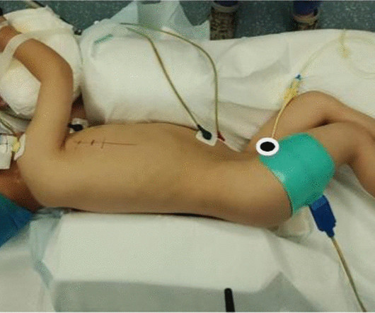
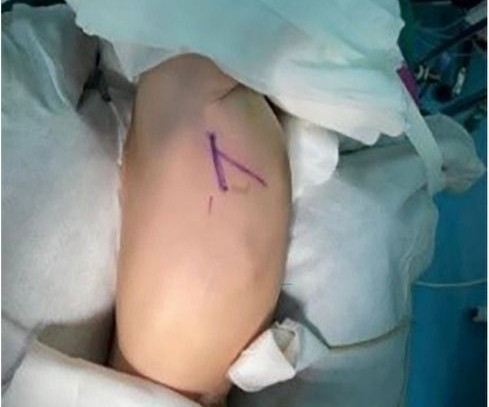
Perimembranous VSD is the commonest type of ventricular septal defect noted in children. Large VSDs can be associated with heart failure in infancy and may need surgery. Device closure is not that well established in perimembranous VSD as in case of muscular VSD.
To compare the therapeutic effects of right vertical infra-axillary thoracotomy (RVIAT) and Standard Median Sternotomy (SMS) in the repair of atrial septal defect (ASD) and ventricular septal defect (VSD), and.
We hypothesize that applying artificial intelligence (AI) to chest x-rays (CXRs) could identify the future risk of PAH in patients with ventricular septal defect (VSD). Methods A total of 831 VSD patients (161 PAH-VSD, 670 nonPAH-VSD) was retrospectively included.
While ventricular septal defects (VSDs) have been linked to an increased risk of infective endocarditis, cases of acquired VSD. Infective endocarditis remains a deadly disease with a significant mortality rate.
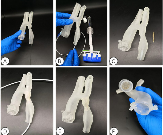
HeartX's portfolio includes advanced products like the JOVE VB Stent for Sinus Venous ASD, JOVE Versatile ASD (VASO), Fenestrated VASO and JOVE PFO, with ongoing developments in PDA and VSD closures as well as advanced visualization systems for structural interventions.
The following are key points to remember from a clinical consensus statement on ventricular septal defect (VSD) complicating acute myocardial infarction (MI).
Patients with pulmonary atresia and ventricular septal defect (PA/VSD) are prone to progressive aortic dilation. However, there are relatively few reports of progressive development of aortic aneurysm or aorti.
Introduction:Ventricular septal defect (VSD) is the most common pediatric defect which benefits from closure at an early age. and excluded patients with VSD diameter >10mm and those with other congenital cardiac anomalies.Results:We included 40 patients with a mean age of 18.95 ± 13.02 The mean VSD size was 6.01 ± 2.29
In repaired tetralogy of Fallot (rTOF), the septal anatomical isthmuses (AI), 3, between the ventricular septal defect (VSD) and pulmonary annulus, and 4, between the VSD and tricuspid annulus, are important ventricular tachycardia (VT) substrates when slow conducting.
One case complicated with ventricular septal defect (VSD). Two patients with PDA and VSD underwent interventional occlusion at the same time without shunt. There were 6 males and 4 females whose average age was (27.6813.45) years. The operation simulation was performed before operation to determine the best operation plan.
Ventricular septal defect (VSD) is the most common congenital cardiac malformation, accounting for approximately 30% of congenital heart defects. Conventional surgical repair using cardiopulmonary bypass is in.
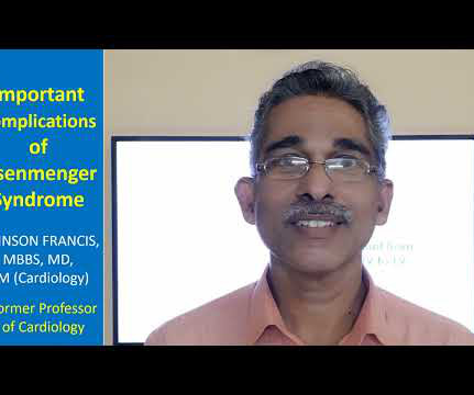
Diagrammatic representation of VSD Eisenmenger. The other Eisenmenger syndromes are not called Eisenmenger complex, only VSD Eisenmenger is called Eisenmenger complex. Large ASDs usually develop Eisenmenger syndrome, may be after decades, not like early development of Eisenmenger syndrome in VSD and PDA.
Bipolar ablation carries a risk of steam pops, conduction system injury, etc but iatrogenic VSD has not previously been described Bipolar radiofrequency (RF) ablation is an adjunctive tool for VT ablation to treat deep intraseptal or mid-myocardial sources of arrhythmias.
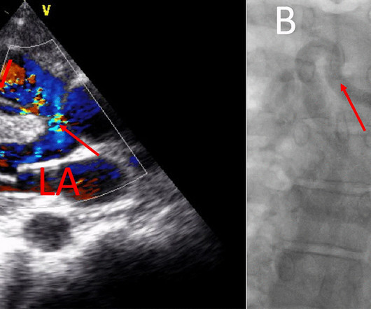
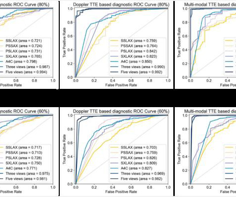
After the child completed the TTE examination, Xie’s auxiliary CHD diagnostic system automatically analyzed the TTE images and computed the probability of each subject being normal, having ASD, or having VSD.
In VSD Eisenmenger and PDA Eisenmenger, you would not expect right atrial enlargement. In VSD and PDA Eisenmenger, the heart size decreases when pulmonary hypertension develops, due to decrease in the shunt. In ASD, shunt is decreasing in Eisenmenger but right atrial enlargement is a component for cardiomegaly.
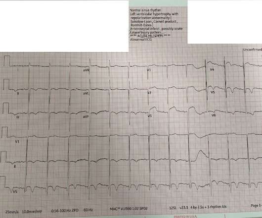
The sign has been described in VSD with biventricular hypertrophy in children. It can be seen with isolated VSD as well as complex ventricular septal defect. Katz-Wachtel Phenomenon or Sign on ECG in VSD An ECG with Katz-Wachtel phenomenon is shown here.
He was diagnosed with a butterfly vertebrae, kidney fullness, sacral dimple, and several heart defects (right aortic arch, VSD, and ASD). He spent 6 days there where we were told he was a candidate for VACTERL.
Introduction Major advances in reperfusion therapies after acute myocardial infarction (AMI) have resulted in dramatic reductions in the rate of mechanical complications including ventricular septal defect (VSD), free wall rupture (FWR) and papillary muscle rupture (PMR).
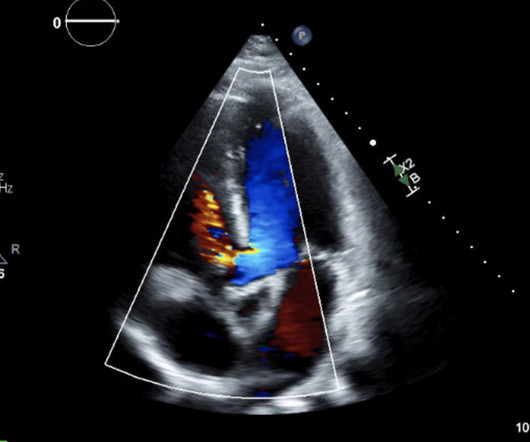
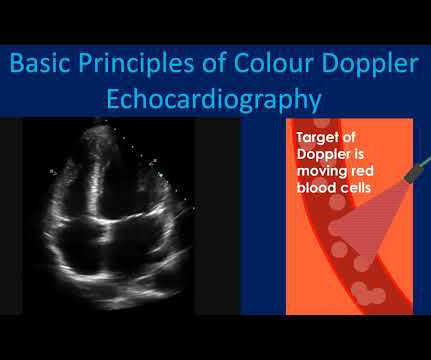
For example, when the VSD is small, the pressure difference between the right and left ventricles is high. Colour Doppler echocardiography is very useful in giving a quick visual assessment of regurgitation and stenosis of heart valves. It will also show abnormal flows as in an atrial or ventricular septal defect.
Curious minds might ask, can’t we decompress LV it self by creating a small VSD. This is to create a small regulatory orifice in the IAS ( A complicated term for a small ASD ) to decompress the LA and reduce pulmonary congestive symptoms. Probably in the thin membranous area.
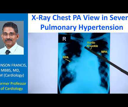
The VSD is partly overrided by the aorta. I am sure that most of you are familiar with echocardiography would have come at the diagnosis and differential diagnosis by this time because it is a very simple view. And this is a better annotated view and I am showing the ventricular septal defect here. Right ventricle, left ventricle.
But in a VSD with pulmonary hypertension A wave is not prominent. It will not occur in the presence of a large VSD which equalizes both right ventricular and left ventricular pressures. Right atrial hypertrophy as in tricuspid stenosis, pulmonary stenosis and pulmonary hypertension.
In addition, the cost of open-heart surgery remains beyond the scope of 90% of the population, ranging from US$6,000 for ventricular septal defect (VSD) closure to US$10,000 for double valve replacement.
Ventricular Septal Defect (VSD) : A hole in the wall (septum) between the heart’s two lower chambers (ventricles). Some of the most common types include: Atrial Septal Defect (ASD) : A hole in the wall (septum) between the heart’s two upper chambers (atria).
Free wall rupture, VSD, Dresslers Syndrome, chronic CHF, anatomic LV aneurysm, LV thrombus, stroke, etc). No further echocardiograms were available after cath. The full thickness infarction with LV aneurysm morphology places him at a higher risk for short and long term complications (e.g., Teaching points: 1.
BackgroundLimited study has shown whether NT-proBNP is related to the prognosis of children wth ventricular septal defect (VSD) surgery. The short- and mid-term clinical outcomes were recorded.
He was transferred to a childrens hospital in Fort Worth, Texas, where his diagnoses were confirmedCoarctation of the aorta, a hypoplastic aortic valve, a large VSD (ventricular septal defect), and an ASD (atrial septal defect). The surgery was a success, and Christopher began to thrive, although complications arose.
This R=S pattern of tall RS complexes brings to mind the Katz-Wachtel phenomenon described in pediatric patients in which the finding of biphasic RS complexes of 50 mm in mid-chest leads V2, V3 or V4 suggests biventricular hypertrophy, especially in children with VSD ( V entricular S eptal D efect ).
We organize all of the trending information in your field so you don't have to. Join thousands of users and stay up to date on the latest articles your peers are reading.
You know about us, now we want to get to know you!
Let's personalize your content
Let's get even more personalized
We recognize your account from another site in our network, please click 'Send Email' below to continue with verifying your account and setting a password.


Let's personalize your content