This site uses cookies to improve your experience. To help us insure we adhere to various privacy regulations, please select your country/region of residence. If you do not select a country, we will assume you are from the United States. Select your Cookie Settings or view our Privacy Policy and Terms of Use.
Cookie Settings
Cookies and similar technologies are used on this website for proper function of the website, for tracking performance analytics and for marketing purposes. We and some of our third-party providers may use cookie data for various purposes. Please review the cookie settings below and choose your preference.
Used for the proper function of the website
Used for monitoring website traffic and interactions
Cookie Settings
Cookies and similar technologies are used on this website for proper function of the website, for tracking performance analytics and for marketing purposes. We and some of our third-party providers may use cookie data for various purposes. Please review the cookie settings below and choose your preference.
Strictly Necessary: Used for the proper function of the website
Performance/Analytics: Used for monitoring website traffic and interactions
Acute extensive portal venous system thrombosis (PVST) can cause lethal complications. Herein, we have for the first time reported the use of anticoagulation combined with systemic thrombolysis by tenecteplase in a male patient with a diagnosis of acute extensive PVST but without liver cirrhosis.
BACKGROUND:Prior clinical trials have demonstrated the efficacy of ultrasound-facilitated catheter-directed thrombolysis (USCDT) for the treatment of acute intermediate-risk pulmonary embolism (PE) using reduced thrombolytic doses and shorter infusion durations. Circulation: Cardiovascular Interventions, Ahead of Print.
To compare the treatment outcomes among percutaneous mechanical thrombectomy (PMT) with AngioJet, Catheter-directed thrombolysis (CDT), and a combination of both.
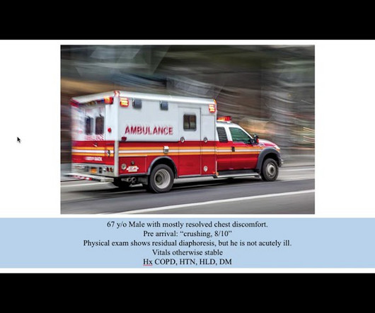
Coronary thrombosis is a dynamic process of platelet aggregation and subsequent coagulation. During spontaneous reperfusion -- whether via thrombolysis, or recruitment of collateral circulation -- there exists characteristic ST/T changes on the ECG. Case Review: [link]
Subjects were divided into "intravenous thrombolysis group" and "non-intravenous thrombolysis group". Result:A total of 1971 patients (437 thrombolysis) were included. In the external validation set, the AUC-ROC values of "PAIST Scale" were 0.855 in the non-thrombolysis group and 0.778 in the thrombolysis group.
Ultrasound showed no thrombosis in the veins of both lower limbs. The patient's father and grandfather have a history of lower limb venous thrombosis. The patient was treated with heparin anticoagulant therapy, catheter thrombus aspiration, and catheter thrombolysis. 76+2_76+3del) gene is extremely rare.
vs. 6.1%, p = 0.0002), deep vein thrombosis (7.3 Patients receiving thrombolysis prior to thrombectomy were less likely to have a prolonged LOS (27.8% p = 0.0002), not receiving IV thrombolysis (OR 2.3, vs. 5.7%, p = 0.0200), intubation for the procedure (86.7% vs. 69.3%, p = 0.0001), complications including pneumonia (18.5%
How common is thrombosis in the culprit artery of Wellen syndrome ? However by no means, we can say thrombosis do not occur. RCA and LCX Wellens do occur, making this entity’s perceived unique importance less certain 3. It is generally believed it is more of a mechanical plaque lesion. Reference 1.
L), intravenous thrombolysis was associated with a reduced number of thrombectomy device passes.Conclusions:Fibrinogen plays a significant role in facilitating thrombosis and the occurrence of LVOS in patients with ICAS. Among patients with lower fibrinogen levels (< 3.2g/L),
Intravenous thrombolysis was deferred as the patient was on pre‐admission anticoagulants. Computed tomography angiography (CTA) showed bilateral vertebral artery occlusion consistent with a history of dissection in the setting of cervical manipulation. Admission National Institutes of Health Stroke Scale (NIHSS) 20.
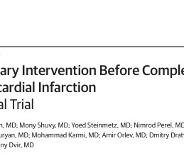
Permanently deferred PCI is other wise called medical management , is practiced by some inferior cardiologists or GPs who never refer such patients to higher centers after a stand alone thrombolysis) * The FFR, iFR RFR, related stuff What if if we are completely blinded to the status of Non IRA vessel ? What do you mean ?
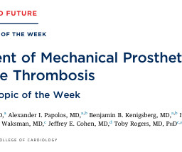
It is to be noted if the obstruction is due to pannus , risk of thrombosis is almost nil and safety of prosthetic balloon valvuloplasty is almost ensured.(Of Generally, overestimation risk of bleeding viz a viz with life threatening thrombosis is quiet common especially in patients with prosthetic valve. I believe, in the above case.
The commonest causes of MINOCA include: atherosclerotic causes such as plaque rupture or erosion with spontaneous thrombolysis, and non-atherosclerotic causes such as coronary vasospasm (sometimes called variant angina or Prinzmetal's angina), coronary embolism or thrombosis, possibly microvascular dysfunction.
This has been termed a “STEMI equivalent” and included in STEMI guidelines, suggesting this patient should receive dual anti-platelets, heparin and immediate cath lab activation–or thrombolysis in centres where cath lab is not available.
There were two self-resolving arterial dissections, one sub-occlusive arterial thrombosis (resolved with 6 weeks of enoxaparin), and one occlusive arterial thrombosis (resolved with alteplase thrombolysis and 6 weeks of enoxaparin). The median fluoroscopy time was 26.1 min min (IQR, 19.2–34.8). Median follow-up was 11.7
A second 12 Lead ECG was recorded: This is a testament to the dynamic nature of coronary thrombosis and thrombolysis. The patient verbalized spontaneous improvement just before 324mg ASA administration. But the lesion is still active!
For this analysis, ACO was defined as angiographic evidence of coronary thrombosis with peak cardiac troponin-I (cTn-I) at least 10 ng/mL or cTn-T ≥ 1 ng/ mL. Blinded physicians adjudicated angiogram reports for coronary lesions and thrombolysis in myocardial infarction (TIMI) flow score.
About Pulmonary Embolism PE is often caused by blood clots in the legs, otherwise known as deep vein thrombosis, that travel through the veins and into the lungs. PE affects around 900,000 people in the U.S. each year, with 10-30% dying within one month of diagnosis.
The challenge we face is whether low-molecular-weight heparin (LMWH) should be used during dialysis.Case presentationA 72-year-old man with a history of hemodialysis for 2 years and 7 months sought medical attention due to thrombosis of the dialysis catheter. The lesion was salvaged via urokinase thrombolysis.
We organize all of the trending information in your field so you don't have to. Join thousands of users and stay up to date on the latest articles your peers are reading.
You know about us, now we want to get to know you!
Let's personalize your content
Let's get even more personalized
We recognize your account from another site in our network, please click 'Send Email' below to continue with verifying your account and setting a password.


Let's personalize your content