This site uses cookies to improve your experience. To help us insure we adhere to various privacy regulations, please select your country/region of residence. If you do not select a country, we will assume you are from the United States. Select your Cookie Settings or view our Privacy Policy and Terms of Use.
Cookie Settings
Cookies and similar technologies are used on this website for proper function of the website, for tracking performance analytics and for marketing purposes. We and some of our third-party providers may use cookie data for various purposes. Please review the cookie settings below and choose your preference.
Used for the proper function of the website
Used for monitoring website traffic and interactions
Cookie Settings
Cookies and similar technologies are used on this website for proper function of the website, for tracking performance analytics and for marketing purposes. We and some of our third-party providers may use cookie data for various purposes. Please review the cookie settings below and choose your preference.
Strictly Necessary: Used for the proper function of the website
Performance/Analytics: Used for monitoring website traffic and interactions
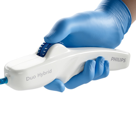
and an investigator in the VIVID study , which contributed to the device’s FDA approval – successfully used the Duo Venous Stent System for the first time outside of a clinical trial. Duo Hybrid has a distinct integrated design that combines multiple zones of differing mechanical properties into a single stent [3].
PTA+DCB, P Key findings include: · One-year primary patency (freedom from both clinically driven target lesion revascularization and duplex ultrasound-derived restenosis) did not differ between groups, despite the significant difference in baseline calcification. · DA+DCB versus 5.9% versus 21.1%, P =0.014). 3 · One-year rates of TLR (16.6%
BACKGROUND:In patients with post-thrombotic syndrome, stent recanalization of iliofemoral veins or the inferior vena cava can restore venous patency and improve functional outcomes. The risk of stentthrombosis is particularly increased during the first 6 months after intervention.
Fortunately, this operator used intravascular ultrasound (IVUS). The operator documented thoughtful consideration of risks and benefits of stent placement. Technically, there was a very narrow landing zone for the stent, and missing this could result in "jailing" the LCx, which is ideally avoided.
The commonest causes of MINOCA include: atherosclerotic causes such as plaque rupture or erosion with spontaneous thrombolysis, and non-atherosclerotic causes such as coronary vasospasm (sometimes called variant angina or Prinzmetal's angina), coronary embolism or thrombosis, possibly microvascular dysfunction.
Stone, MD Mount Sinai Health System tim.hodson Wed, 04/02/2025 - 15:26 March 31, 2025 Using intravascular imaging (IVI) to guide stent implantation during complex stenting procedures is safer and more effective for patients with severely calcified coronary artery disease than conventional angiography, the more commonly used technique.
The secondary outcomes included ischemia-driven target lesion revascularization, target vessel myocardial infarction, death, cardiac death, target vessel revascularization, stentthrombosis, and major adverse cardiac events. OCT was associated with a significant reduction of stentthrombosis compared with ICA (OR, 0.49 [95% CI, 0.26–0.92])
We aimed ultrasound-guided punctures in the proximal two-thirds of axillary arteries with diameters ≥2 mm to insert 7 cm/4 Fr short introducers. Overall, 27/36 procedures were interventional, including 6 aortic valvuloplasties, 6 balloon angioplasties, and 15 stenting procedures. We administrated intra-arterial verapamil (1.25 mg)
His ED cardiac ultrasound (which is not at all ideal for detecting wall motion abnormalities, and is also very operator dependent for this finding) was significant for depressed global EF. It was thought to be an in stent restenosis and thrombosis from a DES placed in the same region 6 months prior.
Deep Vein Thrombosis (DVT) : A blood clot occurring in a deep vein. Causes include infection, malignancy, surgery, scar tissue formation, trauma, deep vein thrombosis (DVT), radiation or other cancer treatment. Carotid Ultrasound : Evaluates blood flow to the brain and detects stroke risk factors.
0.85, P = 0.004), target vessel revascularization (TVR) (P = 0.01), target lesion revascularization (TLR) (P = 0.03) and stentthrombosis (ST) (P = 0.002) in the experimental group (IVUS-guidance) was lower than that in the control group (non-IVUS-guidance). The results showed that the incidence of MACE (RR: 0.63, 95% CI: 0.49–0.82,
We organize all of the trending information in your field so you don't have to. Join thousands of users and stay up to date on the latest articles your peers are reading.
You know about us, now we want to get to know you!
Let's personalize your content
Let's get even more personalized
We recognize your account from another site in our network, please click 'Send Email' below to continue with verifying your account and setting a password.


Let's personalize your content