This site uses cookies to improve your experience. To help us insure we adhere to various privacy regulations, please select your country/region of residence. If you do not select a country, we will assume you are from the United States. Select your Cookie Settings or view our Privacy Policy and Terms of Use.
Cookie Settings
Cookies and similar technologies are used on this website for proper function of the website, for tracking performance analytics and for marketing purposes. We and some of our third-party providers may use cookie data for various purposes. Please review the cookie settings below and choose your preference.
Used for the proper function of the website
Used for monitoring website traffic and interactions
Cookie Settings
Cookies and similar technologies are used on this website for proper function of the website, for tracking performance analytics and for marketing purposes. We and some of our third-party providers may use cookie data for various purposes. Please review the cookie settings below and choose your preference.
Strictly Necessary: Used for the proper function of the website
Performance/Analytics: Used for monitoring website traffic and interactions
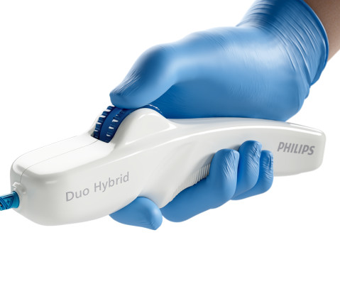
and an investigator in the VIVID study , which contributed to the device’s FDA approval – successfully used the Duo Venous Stent System for the first time outside of a clinical trial. Duo Hybrid has a distinct integrated design that combines multiple zones of differing mechanical properties into a single stent [3].
With IVUS, a tiny ultrasound device is inserted into the vessel via a catheter to produce cross-sectional and 3D images of the vessel for more accurate information on vessel dimensions and plaque morphology. The balloons used in the study were also coated with drugs that help to prevent further plaque buildup. and Korea United Pharm.
PTA+DCB, P Key findings include: · One-year primary patency (freedom from both clinically driven target lesion revascularization and duplex ultrasound-derived restenosis) did not differ between groups, despite the significant difference in baseline calcification. · DA+DCB versus 5.9% versus 21.1%, P =0.014). 3 · One-year rates of TLR (16.6%
Cardiovascular Ultrasound 7. TEVAR Stent Grafts 9. Here are links to the Top 10 viewed DAIC comparison charts from 2023: 1. Guidewires 2. Angiography Systems 3. Hemodynamic Monitoring Systems 4. Cardiac CT Systems 5. Drug-Coated Balloons 6. Echocardiology Reporting Systems 8. Cardiovascular Information Systems (CVIS) 10. ECG Systems
Coronary Intravascular Ultrasound (IVUS) equipment consists of an IVUS catheter, pullback device and the imaging console. IVUS Measurements Measurements include the measurement of lumen, plaque, calcium, remodeling, stent length and volumetric measurements. Incomplete stent apposition can be detected by intravascular ultrasound.
In this week’s View, Dr. Eagle looks at the difference between quantitative coronary angiography versus intervascular ultrasound to guide PCI. He then discusses paclitaxel-coated balloon catheters vs uncoated balloon angioplasty for treating coronary in-stent restenosis.
It is of an elderly woman who complained of shortness of breath and had a recent stent placed. What I had not told him before he made that judgement is that the patient also had ultrasound B-lines of pulmonary edema. Also, we know the patient had a stent. A few days before that, she had had an LAD stent for LAD occlusion.
BACKGROUND:In patients with post-thrombotic syndrome, stent recanalization of iliofemoral veins or the inferior vena cava can restore venous patency and improve functional outcomes. The risk of stent thrombosis is particularly increased during the first 6 months after intervention.
A male in his 40's who had been discharged 6 hours prior after stenting of an inferoposterior STEMI had sudden severe SOB at home 2 hours prior to calling 911. He had diffuse crackles on exam and B-lines on chest ultrasound, and chest x-ray also confirmed pulmonary edema. He had no chest pain.
A man in his mid 60s with history of CAD and stents experienced sudden onset epigastric abdominal pain radiating up into his chest at home, waking him from sleep. Gallbladder ultrasound was negative for stones. It is stented with good angiographic result. See it again now, along with our new Queen of Hearts functionality.
We report a case of TRAD in the early postoperative period, which was successfully managed with intravascular ultrasound-assisted endovascular intervention.Case presentationA 38-year-old man underwent HLA-compatible living kidney transplantation. The transplant renal artery lesion was intervened with a stent.
During the roundtable, participants highlighted the potential of IVUS in guiding revascularization procedures, such as angioplasty and stenting, to optimize outcomes for patients. It provides detailed information about the vessel wall, plaque composition, and blood flow characteristics, enabling more accurate diagnosis and treatment planning.
A bedside cardiac ultrasound performed by a true EM expert (Robert Reardon, who wrote the cardiac ultrasound chapter in Ma and Mateer) showed an inferior wall motion abnormality. The culprit was opened and stented. But there are also new Q-waves, stronly suggesting new infarction.
BACKGROUND:Geographic stent-ostium mismatch is an important predictor of target lesion failure after percutaneous coronary intervention of an aorto-ostial right coronary artery lesion. Optimal visualization of the aorto-ostial plane is crucial for precise stent implantation at the level of the ostium.
Successful PPCI was performed via right femoral artery, with access gained under ultrasound guidance. Additional arterial access via left brachial artery was obtained, and a covered stent was deployed successfully in the right femoral artery with satisfactory haemostasis.
In the rapidly changing field of cardiology, IVUS (Intravascular Ultrasound) and OCT (Optical Coherence Tomography) have seen significant growth. IVUS reveals plaque characteristics, optimal stent usage, and vessel measurements. On the other hand, OCT, with its microscopic accuracy, unveils fine stent positioning and tissue reactions.
BACKGROUND:Bioresorbable scaffolds (BRS) were developed to overcome limitations related to late stent failures of drug-eluting stents, but lumen reductions over time after implantation of BRS have been reported. Six-month angiographic follow-up with optical coherence tomography and intravascular ultrasound was available in 74 patients.
I performed a bedside cardiac ultrasound and the posterior wall appeared to be contracting and shortening normally. Two stents were placed. A posterior ECG was done and showed no ST elevation, not even 0.5 mm in only one posterior lead is highly sensitive and specific for posterior STEMI). The ECG normalized overnight.
On intravascular ultrasound (IVUS), the mid RCA plaque was described as "cratered, inflamed, and bulky," and the OM plaque was described as "bulky with evidence of inflammation and probably ulceration." On the combined basis of angiography and IVUS, this patient received stents to his mid RCA, proximal PDA, and OM.
Meanwhile, over the years, ultrasound moved up from the pelvis, abdomen, right into coronary arteries and heart. Intravascular ultrasound-based interventions are being done in coronary artery, in a few cases to avoid contrast in patients with CKD. (We With zero radiation, MRI came close in the fight with innocuous proton imaging.
Bedside ultrasound with no apparent wall motion abnormalities, no pericardial effusion, no right heart strain. Here are other very interesting posts: Wellens' syndrome: to stent or not? Course : Aspirin 325mg, chemistry, CBC, troponin panel all ordered. Aorta briefly viewed, appears normal caliber and diameter.
Once stabilized, intravascular ultrasound showed significant thrombus and plaque in the LAD. This was treated with a drug-eluting stent, but TIMI 3 flow was not achieved. The patient was placed on an integrilin drip with plans to reevaluate in 24 hours.
Fortunately, this operator used intravascular ultrasound (IVUS). The operator documented thoughtful consideration of risks and benefits of stent placement. Technically, there was a very narrow landing zone for the stent, and missing this could result in "jailing" the LCx, which is ideally avoided.
Under ultrasound guidance, her PT disappeared when the posterior auricular vein collapsed under applied pressure and returned when the pressure was released. Initially, he underwent stent‐assisted coiling of a high‐riding jugular bulb with no change in symptoms. Balloon occlusion test (BOT) of the PCV demonstrated improvement in PT.
He underwent coronary stenting (uncertain which artery). An emergency cardiac ultrasound could be very useful. He underwent immediate CPR, was found to be in ventricular fibrillation, and was successfully resuscitated. I do not have the post-resuscitation ECG. Could this have been avoided?
This was a presumed culprit and a stent was placed. And angiographers tell me that it is sometimes difficult to say for certain based on angiogram alone, without intravascular ultrasound or, better yet, optical coherence tomography. Assuming that was indeed a culprit, then this was ACS.
After guidewire crossing, balloon angioplasty was performed, and a drug-eluting stent was deployed. An intravascular ultrasound was also performed, which was negative for vessel dissection. The left circumflex had 80% proximal stenosis with minimal luminal irregularities in the mid to distal portion.
Two thirds of MINOCA cases are due to atherosclerotic causes One way to prove the diagnosis in this case would have been with intravascular imaging such as optical coherence tomography (OCT) or intravascular ultrasound (IVUS). Fortunately, that is exactly what happened. The patient did well afterward without any recurrence of symptoms.
This was a point of care ultrasound, not a bubble contrast echo. One would not expect wall motion to recover so quickly after stenting, so this is good evidence that the POCUS echo was indeed accurate. What do you think the echocardiogram shows? First trop I returns at 1.5.
A bedside ultrasound revealed a possible anterior wall motion abnormality. There was an LAD occlusion that was opened and stented. The patient was treated with Calcium, Insulin, D50, and bicarbonate, with no change in the ECG. The K returned at 2.9 These T waves are NOT typical for hyperK.
Thirty minutes later the first Troponin I came back elevated at 650 ng/L (normal <26), and bedside ultrasound found anteroseptal akinesia. So the RCA was stented. The patient continued to have chest pain after the RCA was reperfused, so the LAD was then stented. The patient still had chest pain and a third ECG was performed.
It was opened and stented. There was an old ECG for comparison: One year prior with no ST segment abnormalities A bedside cardiac ultrasound was done by the emergency physician. Culprit, stented) 3. A stent was placed and the patient became pain free. Later, the patient was taken to the cath lab. The artery was occluded.
Case continued A bedside ultrasound showed diminished LV EF and of course bradycardia. Angiogram: Culprit Lesion (s): Thrombotic occlusion of the proximal RCA -- stented. When narrow (above His bundle), it is likely to be atropine responsive. 1 mg of Atropine was given and the heart rate increased transiently to 60.
Stone, MD Mount Sinai Health System tim.hodson Wed, 04/02/2025 - 15:26 March 31, 2025 Using intravascular imaging (IVI) to guide stent implantation during complex stenting procedures is safer and more effective for patients with severely calcified coronary artery disease than conventional angiography, the more commonly used technique.
Here are a couple shots with strain, or "speckle tracking" on ED Echo: To, me these look like anterior wall motion abnormality, but I showed them to one of our ultrasound fellows who is very interested in this. It was stented. They read it as normal. She said: This is a tough one. Learning Points : 1. Use the 4-variable formula!!
This is an ultrasound (a bit like the type that we use on pregnant women to look at the baby). An ultrasound will allow you to visualise the heart, measure the sizes of the chambers, assess the heart valves and work out how well the heart functions as a pump.
Smith comment: Point of Care ultrasound is not adequate to rule out wall motion abnormality; moreover, diffuse subendocardial ischemia often has no wall motion abnormality because the epicardium is still contracting. A single DES stent was placed, and the patient did well post-procedure. Academic Emergency Medicine 27(S1): S220.
After many hours, the decided that it was appropriate to do an angiogram and they found a distal LAD occlusion which was opened and stented. This was missed by the physicians, even with a bedside speckle tracking ultrasound: no wall motion abnormality was seen. It was stented. There was no wall motion abnormality. STE60V3 = 2.5
To, me these look like anterior wall motion abnormality, but I showed them to one of our ultrasound fellows who is very interested in this. It was stented. Here are a couple shots with strain, or "speckle tracking" on ED Echo: hyperacute T-waves speckle 1 x4 from Stephen Smith on Vimeo. She said: This is a tough one.
The secondary outcomes included ischemia-driven target lesion revascularization, target vessel myocardial infarction, death, cardiac death, target vessel revascularization, stent thrombosis, and major adverse cardiac events. OCT was associated with a significant reduction of stent thrombosis compared with ICA (OR, 0.49 [95% CI, 0.26–0.92])
Introduction Intravascular ultrasound (IVUS) improves clinical outcome in patients undergoing percutaneous coronary intervention (PCI) but dedicated prospective studies assessing the safety and efficacy of IVUS guidance during primary PCI are lacking. Other endpoints include clinical and procedural outcomes along with post-PCI IVUS findings.
A lower extremity arterial ultrasound revealed elevated velocities in the right proximal superficial femoral artery. Based on these results, Dormu performed a percutaneous transluminal balloon angioplasty and a mechanical atherectomy and stenting of the right superficial femoral artery and stenting of the right superficial femoral artery.
We organize all of the trending information in your field so you don't have to. Join thousands of users and stay up to date on the latest articles your peers are reading.
You know about us, now we want to get to know you!
Let's personalize your content
Let's get even more personalized
We recognize your account from another site in our network, please click 'Send Email' below to continue with verifying your account and setting a password.


Let's personalize your content