This site uses cookies to improve your experience. To help us insure we adhere to various privacy regulations, please select your country/region of residence. If you do not select a country, we will assume you are from the United States. Select your Cookie Settings or view our Privacy Policy and Terms of Use.
Cookie Settings
Cookies and similar technologies are used on this website for proper function of the website, for tracking performance analytics and for marketing purposes. We and some of our third-party providers may use cookie data for various purposes. Please review the cookie settings below and choose your preference.
Used for the proper function of the website
Used for monitoring website traffic and interactions
Cookie Settings
Cookies and similar technologies are used on this website for proper function of the website, for tracking performance analytics and for marketing purposes. We and some of our third-party providers may use cookie data for various purposes. Please review the cookie settings below and choose your preference.
Strictly Necessary: Used for the proper function of the website
Performance/Analytics: Used for monitoring website traffic and interactions
Coronary Intravascular Ultrasound (IVUS) equipment consists of an IVUS catheter, pullback device and the imaging console. IVUS Measurements Measurements include the measurement of lumen, plaque, calcium, remodeling, stent length and volumetric measurements. Incomplete stent apposition can be detected by intravascular ultrasound.
We report a case of TRAD in the early postoperative period, which was successfully managed with intravascular ultrasound-assisted endovascular intervention.Case presentationA 38-year-old man underwent HLA-compatible living kidney transplantation. The transplant renal artery lesion was intervened with a stent.
BACKGROUND:In patients with post-thrombotic syndrome, stent recanalization of iliofemoral veins or the inferior vena cava can restore venous patency and improve functional outcomes. The risk of stent thrombosis is particularly increased during the first 6 months after intervention.
BACKGROUND:Bioresorbable scaffolds (BRS) were developed to overcome limitations related to late stent failures of drug-eluting stents, but lumen reductions over time after implantation of BRS have been reported. Six-month angiographic follow-up with optical coherence tomography and intravascular ultrasound was available in 74 patients.
Bedside ultrasound with no apparent wall motion abnormalities, no pericardial effusion, no right heart strain. Here are other very interesting posts: Wellens' syndrome: to stent or not? Course : Aspirin 325mg, chemistry, CBC, troponin panel all ordered. Aorta briefly viewed, appears normal caliber and diameter.
Or is it a very tight stenosis that does not allow enough flow to perfuse myocardium that has a high oxygen demand from severely elevated BP? This was a presumed culprit and a stent was placed. The T waves in leads II and aVF have deflated, and the T wave in lead III has become terminally negative.
Under ultrasound guidance, her PT disappeared when the posterior auricular vein collapsed under applied pressure and returned when the pressure was released. Initially, he underwent stent‐assisted coiling of a high‐riding jugular bulb with no change in symptoms. A CT venogram revealed left IJ stenosis.
The LAD has diffuse disease with a few areas of moderate stenosis but no flow-limiting lesions. On intravascular ultrasound (IVUS), the mid RCA plaque was described as "cratered, inflamed, and bulky," and the OM plaque was described as "bulky with evidence of inflammation and probably ulceration."
The Queen of Hearts read this ECG as OMI – Low Confidence Click here to sign up for QoH Access The providers taking care of this patient were concerned regarding his clinical history and initial ECG, so they next performed a bedside cardiac ultrasound. The culprit mid LAD lesion was stented.
The left circumflex had 80% proximal stenosis with minimal luminal irregularities in the mid to distal portion. After guidewire crossing, balloon angioplasty was performed, and a drug-eluting stent was deployed. An intravascular ultrasound was also performed, which was negative for vessel dissection.
The cardiologist called this 20% stenosis. Fortunately, this operator used intravascular ultrasound (IVUS). The operator documented thoughtful consideration of risks and benefits of stent placement. However, given the context, she returned for immediate angiography and received a stent to her proximal LAD.
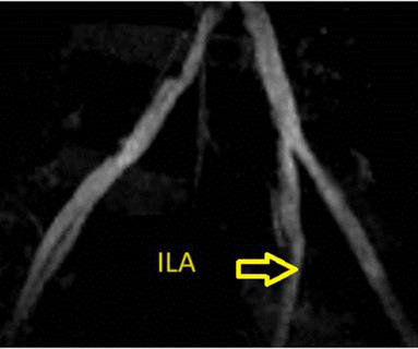
A lower extremity arterial ultrasound revealed elevated velocities in the right proximal superficial femoral artery. Dr. Dormu performed an aortogram of the bilateral lower extremity with bilateral iliac runoff, which revealed a 90% stenosis of the right superficial femoral artery and 100% occlusion of all three tibial vessels.
link] A 62 year old man with a history of hypertension, type 2 diabetes mellitus, and carotid artery stenosis called 911 at 9:30 in the morning with complaint of chest pain. The image on the left shows the LAD before intervention, and the red circled portion on the right indicates the stented region.
It was opened and stented. There was an old ECG for comparison: One year prior with no ST segment abnormalities A bedside cardiac ultrasound was done by the emergency physician. LM: No significant stenosis. LAD: luminal irregularities with a 40% stenosis at the take-off of a D3. D3 has a 95% tubular ostial stenosis.
A middle-aged male with h/o CAD and stents presented with typical chest pressure. A bedside ultrasound was done by the emergency physician, using Speckle Tracking. RBBB does not affect wall motion by dyssynchrony in the way that LBBB does. --Is there likely to be fixed coronary stenosis that led to demand ischemia during pneumonia? --Was
Here are a couple shots with strain, or "speckle tracking" on ED Echo: To, me these look like anterior wall motion abnormality, but I showed them to one of our ultrasound fellows who is very interested in this. It was stented. They read it as normal. She said: This is a tough one. Learning Points : 1. Use the 4-variable formula!!
Angiogram showed a critical LAD thrombotic stenosis. The patient went to cath and had a distal LAD 99% stenosis with thrombus and TIMI-2 flow. After many hours, the decided that it was appropriate to do an angiogram and they found a distal LAD occlusion which was opened and stented. It was stented. He underwent CABG.
To, me these look like anterior wall motion abnormality, but I showed them to one of our ultrasound fellows who is very interested in this. She was treated medically for NonSTEMI, pending next day cath, which showed ulcerated plaque and a 60% thrombotic stenosis in the LAD distal to the first diagonal. It was stented.
They did not have an ultrasound on the ambulance (some local crews are starting to utilize POC limited US in our service areas). He was taken to the cath lab and underwent emergent intervention: Thrombotic stenosis of the proximal RCA (95% with evidence of plaque rupture) is the culprit for the patient's inferoposterior STEMI.
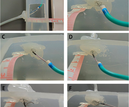
BackgroundPercutaneous coronary intervention (PCI) through the aorto-ostial coronary stent that is protruding into the aorta remains a technical challenge because of the poor coaxial alignment of the guiding catheter and the inability to advance the guidewire into the distal vessel through the stent's central lumen.
We organize all of the trending information in your field so you don't have to. Join thousands of users and stay up to date on the latest articles your peers are reading.
You know about us, now we want to get to know you!
Let's personalize your content
Let's get even more personalized
We recognize your account from another site in our network, please click 'Send Email' below to continue with verifying your account and setting a password.


Let's personalize your content