This site uses cookies to improve your experience. To help us insure we adhere to various privacy regulations, please select your country/region of residence. If you do not select a country, we will assume you are from the United States. Select your Cookie Settings or view our Privacy Policy and Terms of Use.
Cookie Settings
Cookies and similar technologies are used on this website for proper function of the website, for tracking performance analytics and for marketing purposes. We and some of our third-party providers may use cookie data for various purposes. Please review the cookie settings below and choose your preference.
Used for the proper function of the website
Used for monitoring website traffic and interactions
Cookie Settings
Cookies and similar technologies are used on this website for proper function of the website, for tracking performance analytics and for marketing purposes. We and some of our third-party providers may use cookie data for various purposes. Please review the cookie settings below and choose your preference.
Strictly Necessary: Used for the proper function of the website
Performance/Analytics: Used for monitoring website traffic and interactions
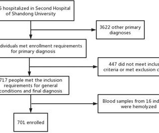
Methods 701 patients were divided into stable coronary artery disease (SCAD), ACS, and control groups. Furthermore, 225 patients who underwent carotid ultrasound were selected from the above 701 patients and were divided into low-risk plaque, medium-to-high risk plaque, and control (without carotid plaques) groups.
In the absence of these factors it is termed spontaneous coronary artery dissection ( SCAD ). At that time the literature suggested: SCAD was rare , Mostly related to pregnancy , Seen on angiography as a dissection flap , and Managed similarly to MI caused by CAD (ASA, BB, lytics/PCI ). The SCAD cases in Lobo et al. Lobo et al.
The ways to tell for certain include intravascular ultrasound (to look for extra-luminal plaque with rupture) or "optical coherence tomography," something I am entirely unfamiliar with. The authors recommend using optical coherence tomography or intravascular ultrasound imaging in patients with evidence of nonobstructive CAD by angiogram.
It was not SCAD (coronary dissection) Highest troponin I was 37,000 ng/L, but it was not measured to peak. And almost all of them could be detected by bedside ultrasound. Conclusion: you may take a few moments to look for dissection with your bedside ultrasound, but when it is a clear STEMI, do NOT waste time with a CT scan.
We organize all of the trending information in your field so you don't have to. Join thousands of users and stay up to date on the latest articles your peers are reading.
You know about us, now we want to get to know you!
Let's personalize your content
Let's get even more personalized
We recognize your account from another site in our network, please click 'Send Email' below to continue with verifying your account and setting a password.


Let's personalize your content