This site uses cookies to improve your experience. To help us insure we adhere to various privacy regulations, please select your country/region of residence. If you do not select a country, we will assume you are from the United States. Select your Cookie Settings or view our Privacy Policy and Terms of Use.
Cookie Settings
Cookies and similar technologies are used on this website for proper function of the website, for tracking performance analytics and for marketing purposes. We and some of our third-party providers may use cookie data for various purposes. Please review the cookie settings below and choose your preference.
Used for the proper function of the website
Used for monitoring website traffic and interactions
Cookie Settings
Cookies and similar technologies are used on this website for proper function of the website, for tracking performance analytics and for marketing purposes. We and some of our third-party providers may use cookie data for various purposes. Please review the cookie settings below and choose your preference.
Strictly Necessary: Used for the proper function of the website
Performance/Analytics: Used for monitoring website traffic and interactions
Objective Early risk assessment of pulmonary arterial hypertension (PAH) in patients with congenital heart disease (CHD) is crucial to ensure timely treatment. We hypothesize that applying artificial intelligence (AI) to chest x-rays (CXRs) could identify the future risk of PAH in patients with ventricular septal defect (VSD).
Patients with pulmonary atresia and ventricular septal defect (PA/VSD) are prone to progressive aortic dilation. However, there are relatively few reports of progressive development of aortic aneurysm or aorti.
In repaired tetralogy of Fallot (rTOF), the septal anatomical isthmuses (AI), 3, between the ventricular septal defect (VSD) and pulmonary annulus, and 4, between the VSD and tricuspid annulus, are important ventricular tachycardia (VT) substrates when slow conducting.
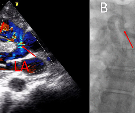
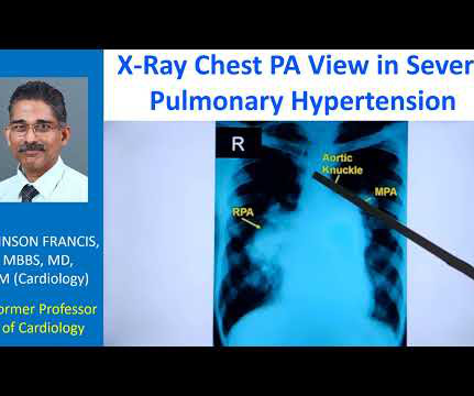
The striking finding is the huge enlargement of the right pulmonary artery, almost aneurysmal dilatation of right pulmonary artery. Main pulmonary artery is also grossly dilated. And you can see left pulmonary artery shadow and rest of it is not seen here. So massive enlargement of pulmonary arteries.
Transcript of the video: Eisenmenger syndrome is an important complication of large left to right shunts which develop later due to development of pulmonary vascular obstructive disease and severe pulmonary hypertension. Diagrammatic representation of VSD Eisenmenger. It is also called Eisenmenger complex. But, leave that alone.
He was diagnosed with a butterfly vertebrae, kidney fullness, sacral dimple, and several heart defects (right aortic arch, VSD, and ASD). However, at the time, we did not know he also had a very rare isolated left pulmonary artery (causing PPHN) as this was hard to detect on the ultrasound and would later be detected at his cath.
Crochetage sign on ECG in ASD ECG in ASD with severe pulmonary hypertension: Tall R’ in V1, ST depression in inferior leads and V2-V5, and T inversion in inferior leads and V1-V6 are seen. The sign has been described in VSD with biventricular hypertrophy in children.
This is to create a small regulatory orifice in the IAS ( A complicated term for a small ASD ) to decompress the LA and reduce pulmonary congestive symptoms. Curious minds might ask, can’t we decompress LV it self by creating a small VSD. Antonio F Corno, et al JTVS 200 3 Remote controlled pulmonary artery banding
The VSD is partly overrided by the aorta. And if it is more than 50% towards the right side, then you think of another condition known as double outlet right ventricle, where both great vessels, aorta and pulmonary artery arises from the right ventricle. You require multiple views to see from where the pulmonary arteries are arising.
Right atrial hypertrophy as in tricuspid stenosis, pulmonary stenosis and pulmonary hypertension. But in a VSD with pulmonary hypertension A wave is not prominent. It will not occur in the presence of a large VSD which equalizes both right ventricular and left ventricular pressures.
We organize all of the trending information in your field so you don't have to. Join thousands of users and stay up to date on the latest articles your peers are reading.
You know about us, now we want to get to know you!
Let's personalize your content
Let's get even more personalized
We recognize your account from another site in our network, please click 'Send Email' below to continue with verifying your account and setting a password.


Let's personalize your content