This site uses cookies to improve your experience. To help us insure we adhere to various privacy regulations, please select your country/region of residence. If you do not select a country, we will assume you are from the United States. Select your Cookie Settings or view our Privacy Policy and Terms of Use.
Cookie Settings
Cookies and similar technologies are used on this website for proper function of the website, for tracking performance analytics and for marketing purposes. We and some of our third-party providers may use cookie data for various purposes. Please review the cookie settings below and choose your preference.
Used for the proper function of the website
Used for monitoring website traffic and interactions
Cookie Settings
Cookies and similar technologies are used on this website for proper function of the website, for tracking performance analytics and for marketing purposes. We and some of our third-party providers may use cookie data for various purposes. Please review the cookie settings below and choose your preference.
Strictly Necessary: Used for the proper function of the website
Performance/Analytics: Used for monitoring website traffic and interactions
Then I always look to see if the initial deflection of the QRS has a lot of voltage change per change in time (seen in tachycardias that are initiated from above the ventricle because the propagate through fast conducting purkinje fiber. Tachycardia exaggerates ST Elevation in LBBB and Paced rhythm 5. Pacemaker mediated tachycardia!
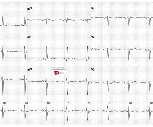
These are typical ECG changes that may indicate a pulmonary embolism. The patient has an acute pulmonary embolism. Sinus tachycardia may be present in acute pulmonary embolism. We see a sinus rhythm with left anterior fascicular block (LAFB) and conspicuous T-wave inversions in the inferior leads and in V1-V6.
Distribution Variance of Focal Atrial Tachycardia Foci and Long-Term Outcomes After Ablation. ABSTRACT Introduction The distribution of the origin of focal atrial tachycardia (FAT) in patients with different ages have not been clearly elucidated. After a mean follow-up of 47.2 months, FAT recurred in 57 patients.
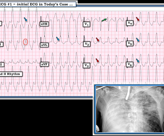
male with pertinent past medical history including Atrial fibrillation, atrial flutter, cardiomyopathy, Pulmonary Embolism, and hypertension presented to the Emergency Department via ambulance for respiratory distress and tachycardia. Bedside ultrasound showed volume depletion and no pulmonary edema. SVT with aberrancy?
He was started on a heparin drip and CTA of the chest was ordered to rule out pulmonary embolism. This is a case like many others posted (see list below) and the EKG from the patient’s original presentation can be quickly recognized as diagnostic for pulmonary embolism. In fact, Kosuge et al. Accessed May 28, 2024.
These are typical ECG changes that may indicate a pulmonary embolism. The patient has an acute pulmonary embolism. Sinus tachycardia may be present in acute pulmonary embolism. Wee see a SR with LAFB and conspicuous T-wave inversions in the inferior leads and in V1-V6. ECG 2 was taken from the same patient 1 year earlier.
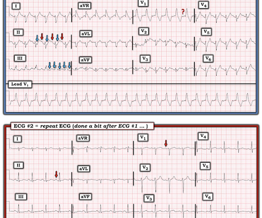
On the monitor patient had wide-complex tachycardia. Differential is ventricular tachycardia versus supraventricular tachycardia with aberrancy versus sinus tachycardia with a aberrancy. After the patient was stabilized with supportive care, the heart rate gradually slowed, confirming sinus tachycardia.
CT of the chest showed no pulmonary embolism but bibasilar infiltrates. Even with tachycardia and a paced QRS duration of ~0.16 (And of course Ken's comments at the bottom) An elderly obese woman with cardiomyopathy, Left bundle branch block, and chronic hypercapnea presented hypoxic with altered mental status. She was intubated.
Electroanatomic mapping guides complex atrial tachycardia ablations; however, challenges may emerge after pulmonary vein isolation. 3D mapping systems can reveal the mechanism of tachycardia and critical areas that need to be ablated.
The ECG in Figure-1 — was obtained from a middle-aged woman with positional tachycardia and diaphoresis with change of position from suprine to sitting. My THOUGHTS on the ECG in Figure-1: The rhythm is sinus tachycardia at ~105/minute ( ie, The R-R interval is regular — and just under 3 large boxes in duration ).
It shows sinus tachycardia with right bundle branch block. Taking a step back , remember that sinus tachycardia is less commonly seen in OMI (except in cases of impending cardiogenic shock). In patients with narrow QRS ( not this patient), this pattern is highly suggestive of acute pulmonary embolism. Both were wrong.
A 77-year-old male with a history of two catheter ablation procedures, including pulmonary vein isolation and superior vena cava (SVC) isolation, presented with symptomatic palpitations. A twelve-lead electrocardiogram revealed atrial tachycardia (AT) with a cycle length of 240 ms.
She was awake, alert, well perfused, with normal mental status and overall unremarkable physical exam except for a regular tachycardia, possible rales at both bases, some mild RUQ abdominal tenderness. Thus, I believe it is a regular, monomorphic, wide complex tachycardia. Or it could simply still be classic VT. What is the Diagnosis?
when the usual negative P wave deflection of sinus tachycardia is nowhere to be found in lead V1 )? While of course possible for the rhythm in ECG #1 to be either AFlutter or fascicular VT — sinus tachycardia immediately becomes a much more likely possibility once we know that this patient is critically ill with multisystem disease.
Introduction Multiple abnormal electrocardiographic findings have been documented in patients experiencing acute pulmonary embolism. Although sinus tachycardia is the most commonly encountered rhythmic disturbance, subsequent reports have highlighted other findings.
There was no pulmonary edema or hypoxia. Here was his initial ECG: Regular Wide Complex Tachycardia. Blood pressure was 180/80. Cardiac Echo showed excellent hyperdynamic function. The computer read the QRS duration as 160 ms. However, many toxins do not show up on tox screens. Sinus --Irregularly irregular?
Epicardial Marshall bundle (MB) are frequently utilized in left atrial tachycardias (LATs) post atrial fibrillation (AF) ablation with pulmonary vein isolation and substrate modification.
Left atrial low-voltage areas (LVAs) are an important arrhythmogenic substrate that can act as an anatomical barrier of reentrant atrial tachycardias (ATs). However, ATs are not always induced in patients with LVAs, and identification of areas with a slow conduction velocity (CV) may help us predict the occurrence of ATs.
Both atria develop from a combination of the primitive atrium, sinus venous, and pulmonary veins.It In all probability, this dilation is a form of atrial tachycardia and atrial cardiomyopathy. In contrast to other tachycardias, with atrial fibrillation (AF), the focus is often speculative, and ablation attempts are made accordingly.
I see the following: There is sinus tachycardia ( upright P wave with fixed PR interval in lead II ) — at the rapid rate of ~130/minute. Sinus Tachycardia and RAD — as already noted above. PEARL # 2: In the absence of associated heart failure ( cardiogenic shock ) — sinus tachycardia is not a common finding in acute MI.
He was in acute distress from pulmonary edema, with a BP of 180/110, pulse 110. He had diffuse crackles on exam and B-lines on chest ultrasound, and chest x-ray also confirmed pulmonary edema. Here is his ED ECG: There is sinus tachycardia. The hypertension alone is the likely etiology of the pulmonary edema.
Ablation of regions demonstrating spatiotemporal dispersion (SD) has been demonstrated as an alternative strategy beyond pulmonary vein isolation in patients with persistent atrial fibrillation. Occurrence of atrial tachycardia (AT) following ablation remains a limitation of this approach.
Particularly in cases of repaired tetralogy of Fallot or double-outlet right ventricle (DORV), a scar resulting from surgery at the right ventricular outflow tract (RVOT) is one of the factors for ventricular tachycardias (VTs).
Pulse field ablation (PFA) using a pentaspline catheter is an effective and safe treatment method for pulmonary vein isolation (PVI) in patients with atrial fibrillation (AF).1,2 We present a case of a macro-reentrant atrial tachycardia in the lateral right atrium (RA) treated by PFA using a pentaspline catheter.
They had already cardioverted at 120 J, then 200 J, which resulted in the following: Ventricular Tachycardia They then cardioverted at 200 J which r esulted in the same narrow complex rhythm shown above, at 185 beats per minute. This would treat both SVT or sinus tachycardia. I suggested esmolol if the heart rate did not improve.
Here is his ECG: Original image, suboptimal quality Quality improved with PM Cardio digitization The ECG is highly suggestive of acute right heart strain, with sinus tachycardia, S1Q3T3, and T wave inversions in anterior and inferior with morphology consistent with acute right heart strain. Moreover, there is tachycardia.
Besides single-shot PFA devices for pulmonary vein isolation (PVI), point-by-point PFA is also gaining importance due to many possible applications. Data on the use is sparse, especially for atrial tachycardia (AT). Pulsed-field ablation (PFA) is becoming increasingly relevant in the field of electrophysiology.
Multifocal Atrial Tachycardia 2. MAT almost always occurs in one of 2 common clinical scenarios : i ) Severe pulmonary disease ( ie, COPD, long-term asthma; pulmonary hypertension ) ; or , ii ) Acutely ill patients with multisystem disease ( ie, patients with sepsis; shock; electrolyte and/or acid-base disorders ).
An Initial ECG was performed: Initial ECG: Sinus tachycardia with prolonged QT interval (QTc of 534 ms by Bazett). She was admitted to the ICU where subsequent ECGs were performed: ECG at 12 hours QTc prolongation, resolution of T wave alternans ECG at 24 hours Sinus tachycardia with normalized QTc interval. No ischemic ST changes.
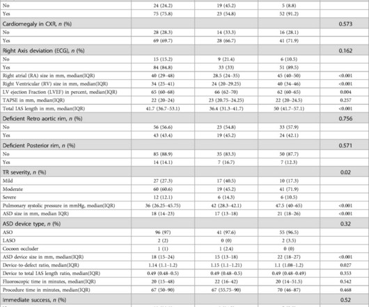
Patients in Group 2 had greater pulmonary artery systolic pressure than those in Group 1 (p-value<0.001). and paroxysmal supraventricular tachycardia (SVT) (5.3%), respectively. The most common symptoms in adults were easy fatigability and dyspnea (63.2% Overall, 88.9% and Group 2: 2.5 (IQR IQR 22.5); P-value=0.111].
BACKGROUND:Varying rates of nonsustained ventricular tachycardia (NSVT) have been reported early after transcatheter pulmonary valve replacement (TPVR) with the Harmony valve, but data regarding rhythm outcomes beyond hospital discharge are limited. Circulation: Cardiovascular Interventions, Ahead of Print.
She had acute pulmonary edema on exam. On arrival, lung ultrasound confirmed pulmonary edema (B lines). Conventional algorithm interpretation: SINUS TACHYCARDIA ABNORMAL RHYTHM ECG Confirmed by over-reading physician Transformed ECG by PM Cardio: PM Cardio interpretation: OMI with Low Confidence Dr.
NOTE: For more on ECG recognition of RVH and/or pulmonary hypertension ( re the qR pattern in lead V1 ) — See ECG Blog #234 and Blog #248. Because of this, it is uncommon to see sinus tachycardia with a prolonged PR interval. This is precisely what we see in Figure-6. Unfortunately — I lack this information.
Methods This study enrolled 233 PeAF patients undergoing pulmonary vein isolation and linear ablation including an MI, roof line, and cavotricuspid isthmus ablation. During the follow-up, 113 (64.9%) RF group patients were free from AF/atrial tachycardia compared to 41 (82.0%) EIVOM group patients (log-rank p = .045). 0.78, p = .006).
She was noted to be in heart failure, with hypotension and tachycardia. Echocardiography demonstrated severe calcific mitral stenosis with pulmonary hypertension. With decompensated heart failure and adequate prior anticoagulation, the patient underwent direct current synchronised cardioversion for her arrhythmia.
The ECG shows sinus tachycardia with RBBB and LAFB, without clear additional superimposed signs of ischemia. The Initial ECG in Today's Case: As per Dr. Meyers — the initial ECG in today's case shows sinus tachycardia with bifascicular block ( = RBBB/LAHB ). Sinus Tachycardia ( common in any trauma patient. ).
Here was his initial ED ECG: There is sinus tachycardia at a rate of about 140 There is profound ST Elevation across all precordial leads, as well as I and aVL. The estimated pulmonary artery systolic pressure is 37 mmHg + RA pressure. QTc was 374, and the formula value was quite high, consistent with LAD occlusion.
Smith interpretation: This is highly likely to be due to extreme right heart strain and is nearly diagnostic of pulmonary embolism. It is of course pulmonary embolism. No d-dimer or CT pulmonary angiogram was done when they discovered that she had normal coronary arteries. Junctional tachycardia? ).
Putting all the findings together; dyspnea, slight tachycardia, delayed R-wave progression, prominent lateral S waves and ST depression maximal where the P waves are largest all point toward pulmonary disease as the cause of the ECG findings. Bedside echo was without wall motion abnormality.
In addition to single-shot devices mainly used for pulmonary vein isolation, focal PFA may provide a treatment option that increases the versatility of the technique. Pulsed field ablation (PFA) has become increasingly important in the treatment of cardiac arrhythmias.
Abstract Introduction It would be helpful in determining ablation strategy if the occurrence of perimitral atrial tachycardia (PMAT) could be predicted in advance. We calculated their E-PMCT.
The ECG and long lead II rhythm strip in Figure-1 — was obtained from a COVID positive patient with persistent tachycardia not responding to Diltiazem. Figure-1: The initial ECG — obtained from a patient with persistent tachycardia. ( Hypoxic injury ( from pneumonia or other acute pulmonary complication ).
We organize all of the trending information in your field so you don't have to. Join thousands of users and stay up to date on the latest articles your peers are reading.
You know about us, now we want to get to know you!
Let's personalize your content
Let's get even more personalized
We recognize your account from another site in our network, please click 'Send Email' below to continue with verifying your account and setting a password.


Let's personalize your content