This site uses cookies to improve your experience. To help us insure we adhere to various privacy regulations, please select your country/region of residence. If you do not select a country, we will assume you are from the United States. Select your Cookie Settings or view our Privacy Policy and Terms of Use.
Cookie Settings
Cookies and similar technologies are used on this website for proper function of the website, for tracking performance analytics and for marketing purposes. We and some of our third-party providers may use cookie data for various purposes. Please review the cookie settings below and choose your preference.
Used for the proper function of the website
Used for monitoring website traffic and interactions
Cookie Settings
Cookies and similar technologies are used on this website for proper function of the website, for tracking performance analytics and for marketing purposes. We and some of our third-party providers may use cookie data for various purposes. Please review the cookie settings below and choose your preference.
Strictly Necessary: Used for the proper function of the website
Performance/Analytics: Used for monitoring website traffic and interactions
IntroductionPercutaneous balloon valvuloplasty is the treatment of choice for critical pulmonary valve stenosis (CPS) and pulmonary valve atresia with intact ventricular septum (PA/IVS) if the ventricle has a suitable size. years), were included. years), were included.
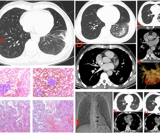
The increased use of radiofrequency ablation (RFA) for atrial fibrillation (AF) has led to a rise in cases of pulmonary vein stenosis or occlusion (PVS/O) as a complication. The actual frequency of pulmonary vein (PV) occlusion remains a topic of debate.
However, the most recurrent cardiac complication in RASopathies is pulmonary valve stenosis (PVS). This has motivated compassionate use of MEK inhibition (MEKi) for rare, but potentially lethal complications such as hypertrophic cardiomyopathy and lymphatic disease.
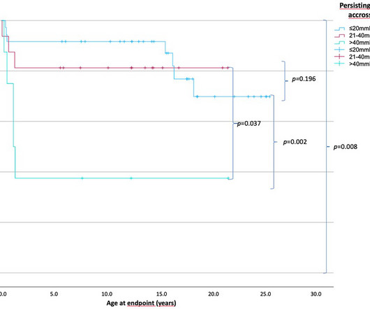
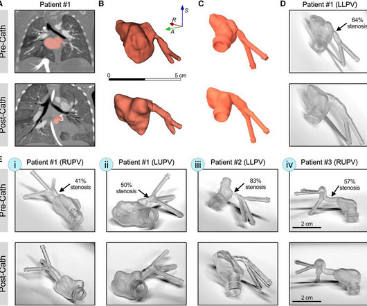
IntroductionPrimary pulmonary vein stenosis (PVS) is a rare congenital heart disease that proves to be a clinical challenge due to the rapidly progressive disease course and high rates of treatment complications. These 3D reconstructions were 3D printed using a clear resin ink and used in a benchtop experimental setup.
Tetralogy of Fallot TOF with pulmonary atresia Pulmonary atresia with intact interventricular septum Tricuspid atresia Double outlet right ventricle Transposition of great arteries with ventricular septal defect and pulmonarystenosis Ebstein’s anomaly of tricuspid valve In DORV and tricuspid atresia, there are also variants with increased pulmonary (..)
Infants with severe valvar pulmonarystenosis (PS) are palliated by transcatheter balloon pulmonary valvuloplasty or surgical pulmonary valvotomy. Either strategy effectively relieves the obstruction, but one long-term sequela is pulmonary regurgitation (PR) necessitating pulmonary valve replacement.
D-Transposition of great arteries Double outlet right ventricle without pulmonarypulmonarystenosis Taussig-Bing anomaly Total anomalous pulmonary venous return Truncus arteriosus Single ventricle (double inlet ventricle, univentricular heart)
Intubated and given nitric oxide for pulmonary hypertension. Four- week echo continues to show pulmonic valve stenosis. Patient scheduled for a balloon valvuloplasty. Unlike adult hearts, the right ventricle comparatively large due to the work it has to do to pump against the high pulmonary pressure before birth.
In the article byTakajoetal, Mortality Patterns in Pediatric Pulmonary Vein Stenosis: Insights Into Right Ventricular Systolic Pressure Associations, which published online on January 17, 2025 (J Am Heart Assoc.2025;2025;14:e037908. Journal of the American Heart Association, Ahead of Print. 2025;2025;14:e037908. DOI:10.1161/JAHA.124.035037)
Some of the late-breaking topics that will be covered include transcatheter aortic valve replacement (TAVR), peripheral artery disease (PAD), and pulmonary embolism (PE): Impact of Age on Procedural Timing for Asymptomatic Severe Aortic Stenosis: Results from the Early TAVR Trial The PERFORMANCE II Trial: A Prospective Multicenter Single Arm Investigation (..)
Such a pattern is consistent with significant left main coronary artery stenosis. Clinical evaluation and X-Ray chest showed features of pulmonary edema. Angiography done after initial stabilization showed severe stenosis of distal left main coronary artery. ST segment elevation is noted in aVR.
In this week’s View, Dr. Eagle looks at the durability of pulmonary vein isolation using pulsed-field ablation, then examines genetic penetrance of dilated cardiomyopathy in genotype-positive relatives.
Cryoablation for atrial fibrillation is a widely used technique for pulmonary vein isolation. Known complications associated with cryoablation include tamponade, phrenic nerve injury, stroke, pulmonary embolism, pulmonary vein stenosis, and atrioesophageal fistulas.
BackgroundRING finger protein 213 (RNF213) p.R4810K is an established risk factor for moyamoya disease and intracranial artery stenosis in East Asian people. Recent evidence suggests its potential association with extracranial cardiovascular diseases, including pulmonary hypertension. All patients had cerebrovascular diseases.
Transcatheter pulmonary valve replacement (TPVR) has become a safe and effective alternative to surgical PVR in tetralogy of Fallot (TOF), isolated pulmonarystenosis (PS), and other congenital heart disease (CHD) variants.
Objective A novel artificial intelligence-based phenotyping approach to stratify patients with severe aortic stenosis (AS) prior to transcatheter aortic valve replacement (TAVR) has been proposed, based on echocardiographic and haemodynamic data. ±15.8 ±15.1 mm Hg, p value: 0.0079).
Voltage mapping was performed before and after PFA operation to help us detect the change in the electrical voltage of the pulmonary veins (PV). 3-Dimensional mapping system showed continuous low potential in the ablation site, and pulmonary vein isolation (PVI) was achieved in all four PV of the patients.
Both atria develop from a combination of the primitive atrium, sinus venous, and pulmonary veins.It Regarding the issue at hand, it is widely known that in cases of mitral stenosis with AF, the left atrium (LA) is larger than the right atrium (RA) due to the obvious reason that the baseline LA was larger at the onset of AF.
Early management of this condition is typically dictated by the degree of pulmonarystenosis (PS) and resulting oxygen saturations. Tetralogy of Fallot (TOF) is the most common form of cyanotic congenital heart disease (CHD).
The CXR demonstrated no pulmonary edema. The LM has an irregular 30% distal stenosis, followed by an 80% ostial LAD stenosis, and total occlusion of the LAD proximally with TIMI grade 1 flow in the distal vessel. The LCX demonstrates an ostial 80% stenosis prior to the bifurcation of a large OM artery. Type II ischemia.
Pulmonary hypertension (PH) is a complex and progressive disorder characterised by elevated pulmonary artery pressure. Transcatheter aortic valve implantation (TAVI) is a minimally invasive surgical procedure that has revolutionised the treatment of severe aortic stenosis (AS).
ABSTRACT Introduction Pulmonary vein (PV) restenosis develops with reported incidence rates of up to 50%. The stenosis was treated with a stent. Balloon angioplasty seems to be the widely preferred treatment of choice. years later he presented with an in-stent restenosis that was successfully treated with a stent-in-stent strategy.
SCAPE is an acronym for sympathetic crash acute pulmonary edema, which can typcially occur in Pickering syndrome with renal artery stenosis [1]. Another term for transient acute pulmonary edema which occurs in renal artery stenosis is flash pulmonary edema. Sympathetic crashing acute pulmonary edem a.
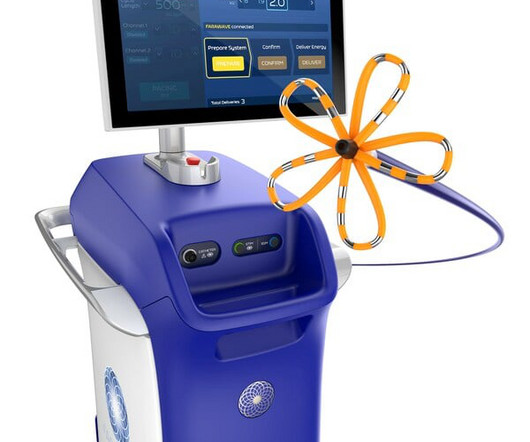
A company statement reported that its PFA System is indicated for the isolation of pulmonary veins in the treatment of drug-refractory, recurrent, symptomatic, paroxysmal (i.e., The FARAPULSE PFA System is indicated for the isolation of pulmonary veins in the treatment of drug-refractory, recurrent, symptomatic, paroxysmal (i.e.,
That is, right ventricle is connecting to aorta, and left ventricle to pulmonary artery. That is, pulmonary artery is transposed over to the right ventricle, and aorta over to the left ventricle, so that normal anatomy is restored. In dextro transposition of great arteries or D-TGA, there is ventriculoarterial discordance.
Clinical introduction A woman in her 30s, a case of rheumatic mitral stenosis status post balloon mitral valvuloplasty 15 years prior, presented to urgent care with palpitations and dyspnoea for 1 week. Echocardiography demonstrated severe calcific mitral stenosis with pulmonary hypertension.
Animal studies have shown that mice with TBX1 gene mutations have smaller left pulmonary arteries compared to wild type mice, defined by a reduced left pulmonary artery (LPA) to right pulmonary artery (RPA) ratio. A single study has shown this translates to humans with 22q11 and structurally normal hearts.
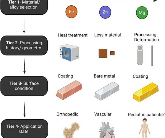
The desired ultimate ability for such devices to treat a vascular stenosis without long-term device-related complications or impeding future treatment continues to evoke excitement in clinicians and engineers alike. The past five years have yielded impressive advancements in fully absorbable metal stent technology.
large ASD, partial anomalous pulmonary venous return, significant tricuspid regurgitation, carcinoid valvular disease, etc,) 2) Conditions causing pressure overload of the RV. Any cause of pulmonary hypertension. There is normal axis, normal R-wave progression in the precordial leads and no intraventricular conduction abnormalities.
Abstract Background The newly introduced nonthermal pulsed field ablation (PFA) is a promising technology to achieve fast pulmonary vein isolation (PVI) with high acute success rates and good safety features. One clinically nonsignificant PV stenosis occurred in the VHPSD group. PFA versus VHPSD for PVI.
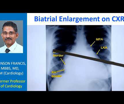
Normally, the main pulmonary artery segment will be concave and left atrial appendage region also will be not prominent. So that is why we see straightening of left border, typically heard of in mitral stenosis with left atrial enlargement and mild pulmonary hypertension. Those are not very clear in this picture.
Chance of precipitating a cyanotic spell are more when pulmonary angiography is attempted through the already narrow right ventricular outflow tract. initial shunt surgery is an option to allow the pulmonary artery branches to grow in size and for a later complete repair of tetralogy of Fallot [1]. If McGoon’s ratio is below 0.8,
According to the analysis of receiver operating characteristic (ROC) curve, AUC, DCA and sensitivity, all seven machine learning models perform well and random forest (RF) machine model was found to perform best (AUC-ROC=0.9008, Accuracy: 0.9008, Precision: 0.6905; Recall: 0.7532, F1: 0.7205).
Stenotic lesions included 16 branch pulmonary arteries, 9 aortic isthmus, 2 right ventricular outflow tracts, and 1 Glenn anastomosis. Percentage of stenosis was 50% (IQR, 36%58%). mm (IQR, 7.59.5) (P<0.001), with median stenosis expansion at 103% (IQR, 51%146%). male) with median age and weight of 3.4 kg (IQR, 9.116.4).
CardioSignal has already developed digital biomarkers for AFib and heart failure, while more solutions could be on the way for aortic stenosis, coronary artery disease, and pulmonary artery hypertension.
In SCAPE (sympathetic crashing acute pulmonary edema), Emergency providers seem now to regularly give high dose NTG, but when the BP is 170/105 in a patient who is not crashing, we often fail to give something to lower afterload. __ Here are some Images: The red circle shows the LAD coursing down the anterior interventricular sulcus.
A transthoracic echocardiogram showed an LV EF of less than 15%, critically severe aortic stenosis , severe LVH , and a small LV cavity. The patient was transported to the CCU for further medical optimization where a pulmonary artery catheter was placed. In fact, bedside ultrasound might even find severe aortic stenosis.
Most popular method is pulmonary vein isolation using these energy sources as the pulmonary veins harbour the triggers for atrial fibrillation. While myocardial tissue is preferentially ablated, effects on adjacent structures like esophagus, phrenic nerve and pulmonary vein tissue is limited with pulsed field ablation.
As clinical presentation is often insidious with nonspecific symptoms, yet morbidity and mortality associated with severe untreated PV disease are significant, a high index of suspicion coupled with appropriate use of imaging techniques is critical in facilitating timely diagnosis and treatment.
BackgroundChronic inflammatory disease (CID) accelerates atherosclerosis and the development of aortic stenosis. Patients with CID were predominantly female (60% versus 44%,P=0.002) and more often had pulmonary disorders (21% versus 13%,P=0.046) and atrial fibrillation (32% versus 20%,P=0.003). and 1.62, respectively).
We organize all of the trending information in your field so you don't have to. Join thousands of users and stay up to date on the latest articles your peers are reading.
You know about us, now we want to get to know you!
Let's personalize your content
Let's get even more personalized
We recognize your account from another site in our network, please click 'Send Email' below to continue with verifying your account and setting a password.


Let's personalize your content