This site uses cookies to improve your experience. To help us insure we adhere to various privacy regulations, please select your country/region of residence. If you do not select a country, we will assume you are from the United States. Select your Cookie Settings or view our Privacy Policy and Terms of Use.
Cookie Settings
Cookies and similar technologies are used on this website for proper function of the website, for tracking performance analytics and for marketing purposes. We and some of our third-party providers may use cookie data for various purposes. Please review the cookie settings below and choose your preference.
Used for the proper function of the website
Used for monitoring website traffic and interactions
Cookie Settings
Cookies and similar technologies are used on this website for proper function of the website, for tracking performance analytics and for marketing purposes. We and some of our third-party providers may use cookie data for various purposes. Please review the cookie settings below and choose your preference.
Strictly Necessary: Used for the proper function of the website
Performance/Analytics: Used for monitoring website traffic and interactions
A male in his 40's who had been discharged 6 hours prior after stenting of an inferoposterior STEMI had sudden severe SOB at home 2 hours prior to calling 911. He was in acute distress from pulmonary edema, with a BP of 180/110, pulse 110. Here is his ED ECG: There is sinus tachycardia. Is this acute STEMI?
The conventional machine algorithm interpreted this ECG as STEMI. It shows sinus tachycardia with right bundle branch block. Taking a step back , remember that sinus tachycardia is less commonly seen in OMI (except in cases of impending cardiogenic shock). In PE, there is almost always some hypoxia without any pulmonary edema.
The prehospital and ED computer interpretation was inferior STEMI: There’s normal sinus rhythm, first degree AV block and RBBB, normal axis and normal voltages. Smith comment: before reading anything else, this case screamed pulmonary embolism to me. The prehospital, ED computer, and final cardiology interpretation was STEMI negative.
So this NSTEMI was likely a STEMI(-)OMI with delayed reperfusion. The patient was admitted as ‘NSTEMI’ which is supposed to represent a non-occlusive MI, but the underlying pathophysiology is analogous to a transient STEMI. See these posts: Chest Pain, ST Elevation, and an Elevated Troponin: Should we Activate the Cath Lab?
Does the ECG represent STEMI-negative OMI findings? Putting all the findings together; dyspnea, slight tachycardia, delayed R-wave progression, prominent lateral S waves and ST depression maximal where the P waves are largest all point toward pulmonary disease as the cause of the ECG findings. How would you mange this patient?
Smith interpretation: This is highly likely to be due to extreme right heart strain and is nearly diagnostic of pulmonary embolism. She was diagnosed with a Non-STEMI and kept overnight for a next day angiogram. Medics recorded the above ECG and called a STEMI alert. It is of course pulmonary embolism.
This certainly looks like an anterior STEMI (proximal LAD occlusion), with STE and hyperacute T-waves (HATW) in V2-V6 and I and aVL. How do you explain the anterior STEMI(+)OMI immediately after ROSC evolving into posterior OMI 30 minutes later? This caused a type 2 anterior STEMI. This prompted cath lab activation.
She had acute pulmonary edema on exam. Prehospital Conventional algorithm interpretation: ANTERIOR INFARCT, STEMI Transformed ECG by PM Cardio: PM Cardio AI Bot interpretation: OMI with High Confidence What do you think? On arrival, lung ultrasound confirmed pulmonary edema (B lines).
A prehospital ECG was recorded (not shown and not seen by me) which was worrisome for STEMI. Here was his initial ED ECG: There is sinus tachycardia at a rate of about 140 There is profound ST Elevation across all precordial leads, as well as I and aVL. The estimated pulmonary artery systolic pressure is 37 mmHg + RA pressure.
This is ischemic ST depression, and could be due to increasing tachycardia, with a heart rate over 130, but that is unlikely given that the patient is now complaining of crushing chest pain and that there was tachycardia all along. Xray was consistent with pulmonary vascular congestion. There is widespread ST depression.
Is this an anterior STEMI with LBBB? Explanation : The patient had a worrisome history: 59 yo with significant substernal chest pressure, so his pretest probability of MI (and even of STEMI) is reasonably high. Additionally, appropriate discordance is common in NonSTEMI, but very unusual in coronary occlusion (STEMI).
The "criteria" for posterior STEMI are 0.5 Is it STEMI or NonSTEMI? The patient had no hypertension, no tachycardia, a normal hemoglobin, no drug use, no hypotension/shock, no murmur of aortic stenosis. Patients with ACS and acute pulmonary edema 3. The troponin I returned at 4.1 mm STE in one lead. This includes: 1.
There is sinus tachycardia. Sinus tachycardia, which exaggerates ST segments and implies that there is another pathology. I have always said that tachycardia should argue against acute MI unless there is cardiogenic shock or 2 simultaneous pathologies. Here is that ECG: What do you think? See Table for diagnostic utility.
No signs for aortic dissection or pulmonary embolus. --"Results were discussed with the ordering physician. If there are T-wave inversions and elevated trops in the context of persistent pain, think of other pathologies such as pulmonary embolism. Thus, Wellens' syndrome should be thought of as a transient OMI or transient STEMI.
A majority of patients with MAT have longstanding pulmonary disease. Rather than antiarrhythmic medication — optimizing pulmonary function is the best treatment approach. Therefore — I’d like to know if this patient had pulmonary problems, a smoking history, or some other significant systemic disorder at the time ECG #3 was obtained.
Clinical Course The paramedic activated a “Code STEMI” alert and transported the patient nearly 50 miles to the closest tertiary medical center. The patient was transported to the CCU for further medical optimization where a pulmonary artery catheter was placed. Look at the aortic outflow tract. What do you see?
In such cases, it is common for tachycardia to exaggerate the ST Elevation And, in fact, there was no new acute MI at this visit - troponins did not rise again. Computer: MI Here is my interpretation: I did not see any NEW acute MI on this ECG This is more consistent with an old MI with persistent ST elevation.
Patients with pulmonary embolism or aortic dissection who have normal variant ST elevation are at high risk of being diagnosed with pericarditis when what they have is far more serious!! normal variant, not pericarditis) A Young Man with Sharp Chest pain (normal variant, not pericarditis) 24 yo woman with chest pain: Is this STEMI?
Bedside ultrasound showed no effusion and moderately decreased LV function, with B-lines of pulmonary edema. Here is his ED ECG: There is obvious infero-posterior STEMI. What are you worried about in addition to his STEMI? See here for management of Polymorphic Ventricular Tachycardia , which includes Torsades.
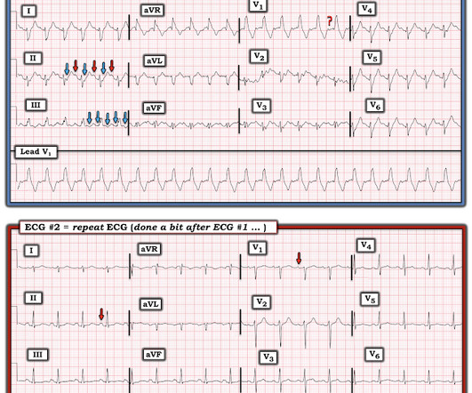
when the usual negative P wave deflection of sinus tachycardia is nowhere to be found in lead V1 )? While of course possible for the rhythm in ECG #1 to be either AFlutter or fascicular VT — sinus tachycardia immediately becomes a much more likely possibility once we know that this patient is critically ill with multisystem disease.
for those of you who do not do Emergency Medicine, ECGs are handed to us without any clinical context) The ECG was read simply as "No STEMI." He was started on a heparin drip and CTA of the chest was ordered to rule out pulmonary embolism. Unfortunately, there was a long wait and the patient left before being seen by a provider.
The morphology of V2-V4 is very specific in my experience for acute right heart strain (which has many potential etiologies, but none more common and important in EM than acute pulmonary embolism). CT angiogram showed extensive saddle pulmonary embolism. He had multiple cardiac arrests with ROSC regained each time.
and the patient was converted to veno-venous (V-V) ECMO due to persistent pulmonary insufficiency. In a series of 18 patients with COVID and ST elevation, 8 were diagnosed with STEMI, 6 of whom had an angiogram and it showed obstructive coronary disease. 12 All STEMI patients had very high cTn typical of STEMI (cTnT > 1.0
When total LM occlusion does present with STE in aVR, there is ALWAYS ST Elevation elsewhere which makes STEMI obvious; in other words, STE is never limited to only aVR but instead it is part of a massive and usually obvious STEMI. All are, however, clearly massive STEMI. This is her ECG: An obvious STEMI, but which artery?
The status of the patients chest pain at this time is unknown : EKG 1, 1300: There is sinus tachycardia and artifact of low and high frequency. However, there is also significant tachycardia , with heart rate of 116, and known hypoxia. Supply-demand mismatch can cause ST Elevation (Type 2 STEMI). An EKG was immediately recorded.
Bedside POCUS showed very poor LV function and a few pulmonary B lines. A Chest X-ray did not show pulmonary edema. This ECG was recorded: It is difficult to appreciate P-waves, but I believe this is sinus tachycardia. This is extremely elevated for a type 2 MI and totally consistent with STEMI. He was put on BiPAP.
The paramedics diagnosis was "Possible Anterolateral STEMI." Chest X-ray also showed pulmonary edema. More proof that a huge STEMI may have normal or near normal initial troponin. Primary VF in this study refers to fibrillation occurring in the absence of shock or pulmonary edema. She was given 2 mg Magnesium.
A 12-lead was recorded, showing "STEMI," but is unavailable. There was never ventricular fibrillation (VF) or ventricular tachycardia (VT), no shockable rhythm. she had severe pulmonary edema. Here is a similar case: Collapse, Ventricular Tachycardia, Cardioverted, Comatose on Arrival. From this site.
We organize all of the trending information in your field so you don't have to. Join thousands of users and stay up to date on the latest articles your peers are reading.
You know about us, now we want to get to know you!
Let's personalize your content
Let's get even more personalized
We recognize your account from another site in our network, please click 'Send Email' below to continue with verifying your account and setting a password.


Let's personalize your content