This site uses cookies to improve your experience. To help us insure we adhere to various privacy regulations, please select your country/region of residence. If you do not select a country, we will assume you are from the United States. Select your Cookie Settings or view our Privacy Policy and Terms of Use.
Cookie Settings
Cookies and similar technologies are used on this website for proper function of the website, for tracking performance analytics and for marketing purposes. We and some of our third-party providers may use cookie data for various purposes. Please review the cookie settings below and choose your preference.
Used for the proper function of the website
Used for monitoring website traffic and interactions
Cookie Settings
Cookies and similar technologies are used on this website for proper function of the website, for tracking performance analytics and for marketing purposes. We and some of our third-party providers may use cookie data for various purposes. Please review the cookie settings below and choose your preference.
Strictly Necessary: Used for the proper function of the website
Performance/Analytics: Used for monitoring website traffic and interactions
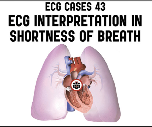
A male in his 40's who had been discharged 6 hours prior after stenting of an inferoposterior STEMI had sudden severe SOB at home 2 hours prior to calling 911. He was in acute distress from pulmonary edema, with a BP of 180/110, pulse 110. Is this acute STEMI? Is this an acute STEMI? -- Unlikely! He had no chest pain.
We discover that for STEMI/OMI vs subendocardial ischemia, we should look for STEMI(-)OMI, subacute OMI, and OMI in the presence of LBBB and RBBB, and consider the differential for diffuse ST depression with reciprocal ST elevation in aVR.
Cardiogenic shock (CS)is the most feared event following STEMI. We tend to perceive CS as an exclusive complication of STEMI. The incidence is half of that of STEMI, i.e., 2.5-5%. might show little elevation with considerable overlap of left main STEMI vs NSTEMI ) 2.Onset ACS pathophysiology is not that simple.
In this ECG Cases blog we look at 10 cases of patients with chest pain, including false positive STEMI, false negative STEMI, and other causes to help hone your ECG interpretation skills in time-sensitive cases where those very ECG skills might save a life.
Background:Little is known about the clinical relevance of interleukin (IL)-6 and the severity of patients with acute ST-elevation myocardial infarction (STEMI). This study examined the possible associations of plasma IL-6 concentrations with TIMI scores in STEMI patients treated with primary percutaneous coronary intervention (PCI).Methods:The
The following ECG was recorded: There is an obvious acute inferior STEMI. Whenever there is inferior STEMI, one should think about Right Ventricular STEMI (RVMI). As 85% of inferior STEMI are due to RCA occlusion [the rest due to occlusion of a "dominant" circumflex (i.e., and STE in lead III > STE in lead II.
So this NSTEMI was likely a STEMI(-)OMI with delayed reperfusion. The patient was admitted as ‘NSTEMI’ which is supposed to represent a non-occlusive MI, but the underlying pathophysiology is analogous to a transient STEMI. See these posts: Chest Pain, ST Elevation, and an Elevated Troponin: Should we Activate the Cath Lab?
The conventional machine algorithm interpreted this ECG as STEMI. In patients with narrow QRS ( not this patient), this pattern is highly suggestive of acute pulmonary embolism. Of course it would also be nice to know about the patients oxygenation: in acute MI there is no hypoxia unless it results in pulmonary edema.
The prehospital and ED computer interpretation was inferior STEMI: There’s normal sinus rhythm, first degree AV block and RBBB, normal axis and normal voltages. Smith comment: before reading anything else, this case screamed pulmonary embolism to me. The prehospital, ED computer, and final cardiology interpretation was STEMI negative.
She had acute pulmonary edema on exam. Prehospital Conventional algorithm interpretation: ANTERIOR INFARCT, STEMI Transformed ECG by PM Cardio: PM Cardio AI Bot interpretation: OMI with High Confidence What do you think? On arrival, lung ultrasound confirmed pulmonary edema (B lines).
This certainly looks like an anterior STEMI (proximal LAD occlusion), with STE and hyperacute T-waves (HATW) in V2-V6 and I and aVL. How do you explain the anterior STEMI(+)OMI immediately after ROSC evolving into posterior OMI 30 minutes later? This caused a type 2 anterior STEMI.
large ASD, partial anomalous pulmonary venous return, significant tricuspid regurgitation, carcinoid valvular disease, etc,) 2) Conditions causing pressure overload of the RV. Any cause of pulmonary hypertension. Troponin T peaked at 2074 ng/L (very high, typical of OMI/STEMI). The LV EF was 57% at formal echo.
In SCAPE (sympathetic crashing acute pulmonary edema), Emergency providers seem now to regularly give high dose NTG, but when the BP is 170/105 in a patient who is not crashing, we often fail to give something to lower afterload. __ Here are some Images: The red circle shows the LAD coursing down the anterior interventricular sulcus.
Smith interpretation: This is highly likely to be due to extreme right heart strain and is nearly diagnostic of pulmonary embolism. She was diagnosed with a Non-STEMI and kept overnight for a next day angiogram. Medics recorded the above ECG and called a STEMI alert. It is of course pulmonary embolism.
Does the ECG represent STEMI-negative OMI findings? Putting all the findings together; dyspnea, slight tachycardia, delayed R-wave progression, prominent lateral S waves and ST depression maximal where the P waves are largest all point toward pulmonary disease as the cause of the ECG findings. How would you mange this patient?
Could this be Septal STEMI (STE in V1 and aVR, with reciprocal ST depression in V4-V6?), In Septal STEMI , transmural ischemia of the septum is recorded by the overlying lead V1 as ST Elevation. Lead III is also on the right and might manifest ST Elevation in Septal STEMI. with ADDED STE in III?
link] Briefly, this woman without significant cardiac history went into pulmonary edema with respiratory failure. In this case, the ECG never mimicked a STEMI. I will proceed to post a couple cases in which SCM does mimic STEMI.
There was some pulmonary edema. This is typical for subendocardial ischemia, not STEMI, and often means left main ischemia or 3 vessel ischemia. Right sided ECGs are best recorded in the context of inferior STEMI. Because of precordial ST depression, clinicians were also worried about posterior STEMI.
To me, this looks like pulmonary edema. B-line predominance bilateral lungs indicates pulmonary edema. B-line predominance bilateral lungs indicates pulmonary edema. 2 months later, he presented in pulmonary edema with atrial flutter and formal echo had EF 20% Why did this happen? It was not a STEMI) 1.
The Non-STEMI, which was an OMI, was diagnosed much faster with AI on the ECG than with troponin. I wonder if this patient had pulmonary disease? The relevance of recognizing pulmonary disease on ECG — is that this may sometimes render assessment for acute OMI more difficult — although this was not the case in today's tracing. (
The "criteria" for posterior STEMI are 0.5 Is it STEMI or NonSTEMI? The patient had been on a long drive, suggesting possible pulmonary embolism (this was unlikely given absence of tachyardia, hypoxia, or any other feature of PE), so we sent a d dimer. Patients with ACS and acute pulmonary edema 3. mm STE in one lead.
This meets "STEMI criteria" However, there is very high voltage, with a very deep S-wave in V2 and tall R-wave in V4. The morphology is not right for STEMI. My interpretation: LVH with secondary ST-T abnormalities, exaggerated by stress, not a STEMI. This is very good evidence that the ST elevation is not due to STEMI.
Right bundle branch block (RBBB) and left anterior fascicular block (LAFB) with very high voltage, consistent with RV hypertrophy (RVH) due to Tetralogy (remember there is a constricted outflow tract of the pulmonary artery, leading to RV hypertrophy.) There is no initial r-wave of the rSR' - there are initial Q-waves.
Is this an anterior STEMI with LBBB? Explanation : The patient had a worrisome history: 59 yo with significant substernal chest pressure, so his pretest probability of MI (and even of STEMI) is reasonably high. Additionally, appropriate discordance is common in NonSTEMI, but very unusual in coronary occlusion (STEMI).
Smith : there is some minimal ST elevation in V2-V6, but does not meet STEMI criteria. Transient STEMI has been studied and many of these patients will re-occlude in the middle of the night. The estimated pulmonary artery systolic pressure is 27 mmHg + RA pressure. Is it normal STE? This is a "Transient OMI".
No signs for aortic dissection or pulmonary embolus. --"Results were discussed with the ordering physician. If there are T-wave inversions and elevated trops in the context of persistent pain, think of other pathologies such as pulmonary embolism. Thus, Wellens' syndrome should be thought of as a transient OMI or transient STEMI.
Without seeing the patient, my interpretation of the first ECG was: likely normal variant ST-elevation (early repolarization), with a small possibility of pericarditis, and almost no possibility of acute coronary occlusion (STEMI). and therefore highly unlikely to be STEMI. Does subsegmental pulmonary embolism matter?
Xray was consistent with pulmonary vascular congestion. 20% of cases that everyone would call a STEMI have a competely open artery by the time of angiogram 60-90 minutes later. Bedside echo showed no evidence of reduced EF, no signs of right heart strain, no regional wall abnormality. Lung exam showed diffuse B lines bilaterally.
In this ECG Cases blog, Jesse McLaren and Rajiv Thavanathan explore how ECG and POCUS complement each other for patients presenting to the emergency department with shortness of breath or chest pain. They explain complementary diagnostic insights into pericardial effusion and cardiac tamponade, occlusion MI and RV strain.
A prehospital ECG was recorded (not shown and not seen by me) which was worrisome for STEMI. A previous ECG from 4 years prior was normal: This looks like an anterior STEMI, but it is complicated by tachycardia (which can greatly elevate ST segments) and by the presentation which is of fever and sepsis.
Was there pulmonary edema? Repeat ECG showing no STEMI, only non-specific ST-segment and T-wave abnormalities, unchanged from prior" Transferred to surgery for exploration but diagnostic studies were too indeterminate to be certain of intra-abdominal pathology. Then they were worried about sepsis as an etiology of hypotension.
A majority of patients with MAT have longstanding pulmonary disease. Rather than antiarrhythmic medication — optimizing pulmonary function is the best treatment approach. Therefore — I’d like to know if this patient had pulmonary problems, a smoking history, or some other significant systemic disorder at the time ECG #3 was obtained.
The medics were worried about STEMI, as it meets STEMI criteria. The troponins are NOT consistent with STEMI (OMI), which typically has a troponin I of at least 5 ng/mL. P.S.: Keep in mind that competing conditions ( ie, hyperkalemia, acute infarction, conduction defects, pulmonary disease ) may mask ECG diagnosis of LVH.
This is a troponin I level that is almost exclusively seen in STEMI. So this is either a case of MINOCA, or a case of Type II STEMI. If the arrest had another etiology (such as old scar), and the ST elevation is due to severe shock, then it is a type II STEMI. I believe the latter (type II STEMI) is most likely.
Clinical Course The paramedic activated a “Code STEMI” alert and transported the patient nearly 50 miles to the closest tertiary medical center. The patient was transported to the CCU for further medical optimization where a pulmonary artery catheter was placed. Look at the aortic outflow tract. What do you see?
Exclusion criteria were age less than 18, SBP less than 100 mmHg, echocardiogram with EF less than 50%, STEMI, pregnancy, and trauma. While sensitivity of this sign is very low — its presence is highly suggestive of longstanding and severe pulmonary disease. Clin Cardiol 22:334-344, 1999 ).
ECG read as: "Shows T wave inversions in the inferior leads and less than 1mm STE in V2, without STEMI criteria." CT pulmonary angiogram was negative for pulmonary embolism. All very very subtle. So the patient was placed back in the waiting room like many others. Aspirin was given. Second troponin T resulted at 1,318 ng/L.
Still does not meet STEMI criteria, but it is an obvious OMI And then another one became more obvious: Cath lab was activated and a 100% RCA occlusion was found. There are many potential causes of ST-T wave abnormalities (ie, LV “strain” from LVH; medication effect; electrolyte disturbance; pulmonary embolus, etc.). Learning Points: 1.
I knew that, if the patient had presented with chest discomfort, that this ECG is diagnostic of inferior posterior OMI, even though it is not a STEMI. The ACC/AHA guidelines mandate less than 2 hours cath for patients with ACS with refractory pain, pulmonary edema, or electrical or hemodynamic instability.
In this ECG Cases blog we look at 6 patients who presented with cardiorespiratory symptoms, possibly from COVID and illustrate the dangers of anchoring, being hypervigilant for cardiovascular complications, and why testing for COVID in patients being admitted for ACS is important.
In this ECG Cases blog we review 10 cases of possible artifact, lead reversal and lead misplacement. Can you spot the abnormalities and avoid the misdiagnosis? The post ECG Cases 29 Misdiagnosis from Lead Misplacement, Artifact and Lead Reversal appeared first on Emergency Medicine Cases.
The limb leads have been removed because there was no ST elevation in those leads, the QRS complexes have been obscured because this is irrelevant to STEMI criteria, and red lines have been added to measure ST segment elevation. But STEMI criteria ignore all this and look at ST segments in isolation.
The computerized interpretation for this tracing was, “Sinus rhythm; Normal ECG” — and attention of acute care providers was apparently focused on attending to this patient’s pulmonary problems. I focus my comments on the first 2 tracings shown in this case — which for clarity, I have put together in Figure-1.
Patients with pulmonary embolism or aortic dissection who have normal variant ST elevation are at high risk of being diagnosed with pericarditis when what they have is far more serious!! normal variant, not pericarditis) A Young Man with Sharp Chest pain (normal variant, not pericarditis) 24 yo woman with chest pain: Is this STEMI?
We organize all of the trending information in your field so you don't have to. Join thousands of users and stay up to date on the latest articles your peers are reading.
You know about us, now we want to get to know you!
Let's personalize your content
Let's get even more personalized
We recognize your account from another site in our network, please click 'Send Email' below to continue with verifying your account and setting a password.


Let's personalize your content