This site uses cookies to improve your experience. To help us insure we adhere to various privacy regulations, please select your country/region of residence. If you do not select a country, we will assume you are from the United States. Select your Cookie Settings or view our Privacy Policy and Terms of Use.
Cookie Settings
Cookies and similar technologies are used on this website for proper function of the website, for tracking performance analytics and for marketing purposes. We and some of our third-party providers may use cookie data for various purposes. Please review the cookie settings below and choose your preference.
Used for the proper function of the website
Used for monitoring website traffic and interactions
Cookie Settings
Cookies and similar technologies are used on this website for proper function of the website, for tracking performance analytics and for marketing purposes. We and some of our third-party providers may use cookie data for various purposes. Please review the cookie settings below and choose your preference.
Strictly Necessary: Used for the proper function of the website
Performance/Analytics: Used for monitoring website traffic and interactions
This case report describes an atypical presentation of CAS in a 68-year-old white British male with cardiovascular riskfactors. Coronary angiography identified moderate stenosis of the right coronary artery (RCA), without significant flow restriction by invasive pressure wire interrogation.
BackgroundThe utility of screening for the degree of common carotid artery (CCA) stenosis as a predictor of cardiovascular disease (CVD) in a general population remains unclear.Methods and ResultsWe studied 4775 Japanese men and women whose CCA was measured using bilateral carotid ultrasonography at baseline (April 1994–August 2001).
About a fifth of all ischemic strokes are attributed to embolization of ruptured atherosclerotic plaque from carotid arterial stenosis. But it has been difficult to predict which person with asymptomatic carotid artery stenosis is likely to progress to symptomatic carotid disease and stroke. J Am Coll Cardiol. 2024.03.389.
BUT — Cardiac catheterization done a little later did not reveal any significant stenosis. Despite the absence of significant coronary stenosis on her post-arrest cath — the ECG in Figure-1 is clearly diagnostic of an extensive anterolateral STEMI ( presumably from acute LAD [ L eft A nterior D escending ] coronary artery occlusion).
Lp(a) is emerging as an important, yet under-recognized, potential riskfactor for cardiovascular disease due to its ability to promote the development of plaques within artery walls, clot formation and aortic valve calcification. The development of the Tina-quant Lipoprotein (a) Gen.2 2022 Aug, 80 (9) 934946 Kronenberg F.
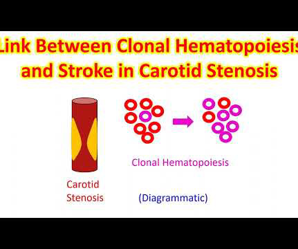
Asymptomatic high-grade carotid stenosis is an important therapeutic target for stroke prevention. Transcarotid artery revascularization has a favorable periprocedural risk profile, but randomized trials comparing it to intensive medical management are lacking.
MINOCA may be due to: coronary spasm, coronary microvascular dysfunction, plaque disruption, spontaneous coronary thrombosis/emboli , and coronary dissection; myocardial disorders, including myocarditis, takotsubo cardiomyopathy, and other cardiomyopathies. There may be a chronic tight stenosis and a non-obstructed lesion that thrombosed.
Incident carotid plaques and their vulnerability were detected by carotid ultrasound at follow-up (2021). Higher sdLDL-C or sdLDL-C/LDL-C ratio, but not LDL-C, was significantly associated with an increased risk of incident carotid plaques. years (SD=0.14). years (SD=0.14). 9.90];P=0.027;Pfor linear trend=0.025).
The scan also showed “scattered coronary artery plaques”. __ Smith comment 1 : the appropriate management at this point is to lower the blood pressure (lower afterload, which increases myocardial oxygen demand). Smith comment : Is the ACS (rupture plaque) with occlusion that is now reperfusing?
Doppler ultrasonography performed a day after the operation showed an increase in systolic blood velocity, with no observed urine output and raising a suspicion of arterial anastomotic stenosis. The transplant renal artery lesion was intervened with a stent.
The vessels with reduced CFR presented a significantly higher prevalence of obstructive CAD (37% vs. 26%; P < 0.001), diffused atherosclerosis (22% vs. 11%; P < 0.001), low-attenuation plaque (6% vs. 3%; P = 0.030), and positive remodeling (7% vs. 2%; P = 0.001). FAI was higher in vessels with reduced CFR (−80.8 HU HU vs. −81.8 HU;
link] A 62 year old man with a history of hypertension, type 2 diabetes mellitus, and carotid artery stenosis called 911 at 9:30 in the morning with complaint of chest pain. Smith's comments in the May 19, 2020 post : — Non-obstructive coronary disease does not ne cessarily imply no plaque rupture with thrombus. It is not rare.
The degree and extent of subclinical coronary atherosclerosis were evaluated by coronary computed tomographic angiography, and ≥50% diameter stenosis was defined as significant. After adjustment for cardiovascular riskfactors, depression was not significantly associated with any coronary plaque (adjusted odds ratio [OR], 1.05 [95% CI, 0.78–1.41];P=0.746),
I quickly reviewed the patient’s records and saw that she was a 53 year old woman with a history of BMI 40, but no other identifiable riskfactors for coronary artery disease. Learning Points: Type 1 MI is the type we are most familiar with: rupture of atherosclerotic plaque with production thrombus or platelet fibrin aggregates.
Nevertheless, the relationship between CAC and the susceptibility of a plaque to provoke a thrombotic event remains incompletely understood. Limited spatial resolution and blooming artifacts may hinder estimation of degree of coronary artery stenosis. This review summarizes the current understanding and literature on CAC.
Given the consistency of the clinical profile with typical angina, associated riskfactors, and abnormal ECG findings, a cardiology consult was promptly requested. Category 1 : Sudden narrowing of a coronary artery due to ACS (plaque rupture with thrombosis and/or downstream showering of platelet-fibrin aggregates. Severe HTN d.
However, CTA head and neck 4 days later demonstrated 90 percent stenosis of the mid left V2 at the C3‐4 level and a 75‐90 percent stenosis of the left mid V2 segment at the C5‐6 level (hard and soft plaque in these areas). He also had moderate stenosis of the right V4 segment.
FFR CT was measured 1 cm distal to the coronary plaque or in the middle of the segments if no coronary lesions were present. Sensitivity analysis was done by cardiac riskfactors, degree of stenosis and image quality. Results A total of 535 coronary segments in 60 patients were assessed.
If the arrest was caused by acute MI due to plaque rupture, then the diagnosis is MINOCA. Here is my comment on MINOCA: "Non-obstructive coronary disease" does not necessarily imply "no plaque rupture with thrombus." They often cannot even be recognized as culprits, as fissured or ulcerated plaque. myocarditis).
24: Joint American College of Cardiology/Journal of the American College of Cardiology Late-Breaking Clinical Trials (Session 402) Saturday, April 6 9:30 – 10:30 a.m.
A completely healthy 30-something year old woman with no cardiac riskfactors had sudden onset of bilateral trapezius pain that radiated around to her throat. R Riskfactors = 0 5. Risk of 30-day adverse events is less than 1.7%. She called 911. mm at the J-point, relative to the PQ junction. A Age: = 0 4.
No thromboembolism risks, not pleuritic, no radiation to the back. No cardiac riskfactors, no cocaine use. It showed a 99% stenosis in the RCA, and proximal to a posterolateral branch. Nevertheless, even young people have atherosclerosis and plaque rupture. History: Onset of CP 2.5 hours prior to ED arrival.
A completely healthy 30-something year old woman with no cardiac riskfactors had sudden onset of bilateral trapezius pain that radiated around to her throat. R Riskfactors = 0 5. Risk of 30-day adverse events is less than 1.7%. The ECG told the story. The first troponin was below the level of detection (LoD).
Coronary computed tomography angiography (CCTA) is routinely used to diagnose CAD caused by the narrowing (stenosis) or blockage of the coronary arteries that supply the heart with blood. More than 8 million Americans visit hospital emergency departments experiencing chest pain every year.
Previous studies demonstrated bilateral symmetry in atherosclerotic plaque burden and calcification scores. Within each artery, the range of slices with wall thickness > 2mm on any scan was included as potential atherosclerotic plaque. Each subject underwent an average of 4.8 scans over 5.2 scans over 5.2
Demographic characteristics, vascular riskfactors, and the results of preoperative serum biochemistry were measured and collected. The riskfactors for vulnerable carotid plaque were analyzed. A Lasso-logistic regression prediction model was developed and compared with traditional logistic regression models.
42% of adults are considered obese , increasing their risk of diabetes, hypertension, and cardiovascular issues. Additionally, 10% of the global population suffers from chronic kidney disease , with diabetes and hypertension as significant riskfactors. In the U.S.,
The therapeutics of coronary stenosis has become a technogical wonder, interwoven with statistical wordplay in the last few decades. Like, viability, scars, futility, and benefits of revascularization, imaging-assisted PCI, impact of PCI on exercise capacity, importance of riskfactor management, etc. 2016 Dec;9(12):e003726.
We organize all of the trending information in your field so you don't have to. Join thousands of users and stay up to date on the latest articles your peers are reading.
You know about us, now we want to get to know you!
Let's personalize your content
Let's get even more personalized
We recognize your account from another site in our network, please click 'Send Email' below to continue with verifying your account and setting a password.


Let's personalize your content