This site uses cookies to improve your experience. To help us insure we adhere to various privacy regulations, please select your country/region of residence. If you do not select a country, we will assume you are from the United States. Select your Cookie Settings or view our Privacy Policy and Terms of Use.
Cookie Settings
Cookies and similar technologies are used on this website for proper function of the website, for tracking performance analytics and for marketing purposes. We and some of our third-party providers may use cookie data for various purposes. Please review the cookie settings below and choose your preference.
Used for the proper function of the website
Used for monitoring website traffic and interactions
Cookie Settings
Cookies and similar technologies are used on this website for proper function of the website, for tracking performance analytics and for marketing purposes. We and some of our third-party providers may use cookie data for various purposes. Please review the cookie settings below and choose your preference.
Strictly Necessary: Used for the proper function of the website
Performance/Analytics: Used for monitoring website traffic and interactions
Pulmonary arterial hypertension is a disease of the pulmonary vasculature, resulting in elevated pressure in the pulmonary arteries and disrupting the physiological coordination between the right heart and the pulmonary circulation. Journal of the American Heart Association, Ahead of Print.
BackgroundDespite the poor outcomes related to the presence of pulmonary hypertension, it often goes undiagnosed in part because of low suspicion and screening tools not being easily accessible such as echocardiography. Each 15second PCG, recorded using a digital stethoscope, was processed to generate 5second melspectrograms.
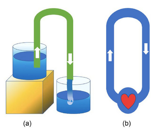
Cardiovascular physiology has long held that the heart is a mechanical pump and that the heart’s propulsive power is the main driver of blood flow throughout the body. While he didn't fully grasp the complete circulatory system, his insights into the pulmonary circulation were notable for the time.
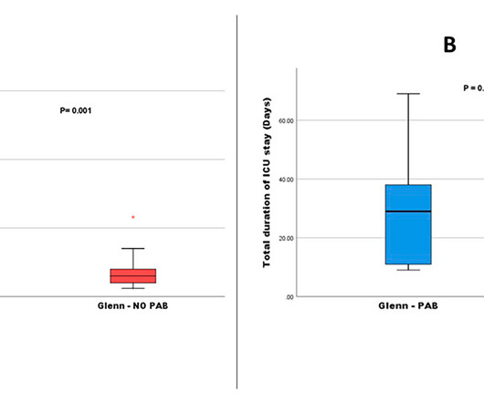
Although pulmonary artery banding (PAB) has been generally acknowledged as an initial palliative treatment for patients having single ventricle (SV) physiology and unrestrictive pulmonary blood flow (UPBF), it.
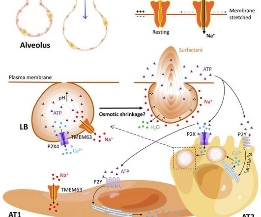
Pulmonary surfactant is a lipoprotein complex lining the alveolar surface to decrease the surface tension and facilitate inspiration. TMEM63A/B were predominantly localized at the limiting membrane of the lamellar body (LB), a lysosome-related organelle that stores pulmonary surfactant and ATP in AT2 cells.
Welcome to the Physiology Friday newsletter. Physiologically Speaking is a reader-supported publication. There’s a long-running debate in exercise physiology about what limits VO2 max. Physiology is integrative, not isolated. To receive new posts and support my work, consider becoming a free or paid subscriber.
Abstract: Angiomotin-like 2 (AMOTL2) is related to numerous physiological and pathological conditions by affecting signal transduction. However, whether AMOTL2 is linked to pulmonary arterial hypertension (PAH) has not been addressed. AMOTL2 was downregulated in hypoxia-stimulated pulmonary arterial smooth muscle cells (PASMCs).
This approach allowed us to evaluate the effect of childhood body size on 11 measures of right heart and pulmonary circulation independent of other anthropometric traits at various stages in the lifecourse. 0.33];P=3×10⁻⁷) independent of adulthood body size.
Challenges in the diagnosis of pulmonary hypertension Pulmonary hypertension (PH) is a clinical–physiological syndrome thought to affect 1% of the global population. 1 PH is defined haemodynamically by mean pulmonary artery pressure >20 mm Hg, resulting in right ventricular (RV) overload and often RV failure.
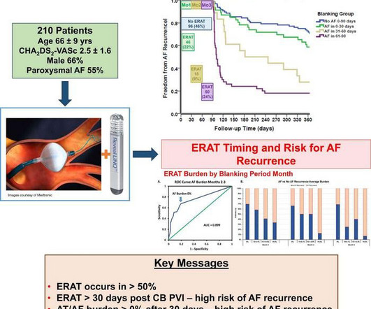
This forms the physiologic rationale for the accepted convention of a three-month blanking period, during which arrhythmia recurrences are presumed to be relatively benign and not indicative of treatment failure. also emphasize the importance of continuous monitoring.
Cardiopulmonary exercise testing is a valuable tool for assessing functional capacity, evaluating cardiac and pulmonary pathology, and providing guidance on prognosis and interventional recommendations.
Extensive ablation, including posterior wall isolation (PWI) is advocated for radiofrequency ablation (RFA) of persistent atrial fibrillation (peAF) to decrease the high recurrence-rates reported when only pulmonary vein isolation (PVI) is performed.1
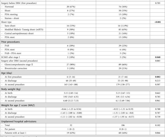
Background:Neonates with complex congenital heart disease and pulmonary overcirculation have been historically treated surgically. 3.3]) underwent a PFR procedure; 15 (88%) had single ventricle physiology; 15 (88%) were high-risk surgical candidates. Circulation: Cardiovascular Interventions, Ahead of Print. kilograms [IQR, 2.1–3.3])
Frailty develops as a result of age-related decline in many physiological systems and is associated with increased vulnerability to adverse outcomes following thoracic surgery. We prospectively tested our hypo.
Moreover, the definitions of elevated exercise pulmonary artery (PA) and PA wedge pressure (PAWP) for this population have not been described. Abstract Background The normal (i.e., expected) hemodynamics in adults post-Fontan remain poorly delineated.
ET (endothelin) is a powerful vasoconstrictor 21-amino acid peptide present in many tissues, which exerts many physiological functions across the body and participates as a mediator in many pathological conditions. Hypertension, Ahead of Print.
link] Briefly, this woman without significant cardiac history went into pulmonary edema with respiratory failure. I refer you to the video case presentation by one of my colleagues, Dr. Rob Reardon (who has, by the way, a fantastic collection of ED ultrasound cases). The apparent trigger was stress from losing custody of children.
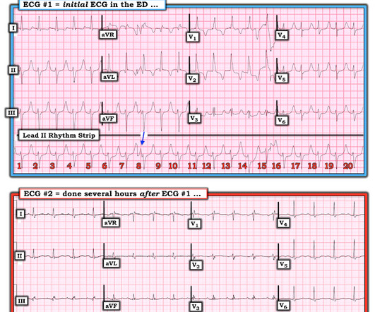
KEY Point: Although true that patients with longstanding, severe pulmonary disease may manifest a QRST complex in standard lead I with marked overall reduction in QRST amplitude ( See ECG Blog #65 — regarding Schamroth’s Sign ) — you should never normally see a completely flat line in any of the standard limb leads.
That is, right ventricle is connecting to aorta, and left ventricle to pulmonary artery. That is, pulmonary artery is transposed over to the right ventricle, and aorta over to the left ventricle, so that normal anatomy is restored. In dextro transposition of great arteries or D-TGA, there is ventriculoarterial discordance.
Notice I did not say "pulmonary embolism," because any form of severe acute right heart strain may produce this ECG. This includes, but is not limited to, PE, asthma/COPD exacerbation, hypoxic vasoconstriction from pneumonia, acute pulmonary hypertension exacerbation. Differences of Pulmonary Embolism T-waves from Wellens' T-waves: 1.
However, stabilization was expected to be temporary due to ongoing physiologic changes of pregnancy. After discharge, she was scheduled for a 2-week postpartum visit including echocardiogram, EKG, and NT-proBNP.Discussion:Given the patient's acute decompensation and fluid overload, medical optimization was essential prior to delivery.
My answer: "This is classic for PE, but it can also be present in any hypoxia due pulmonary hypoxic vasoconstriction and resulting acute pulmonary hypertension and acute right heart strain. The ECG of most patients with longstanding pulmonary disease show more r wave progression than I see in ECG #1. This is NOT Wellens.
This suggests that there is pulmonary hypertension and thus possibly RVH. The estimated pulmonary artery systolic pressure is 31 mmHg + RA pressure. In a patient with RVH — the finding of a qR pattern has been closely correlated with pulmonary hypertension. Right atrial enlargement, severe. Severe tricuspid regurgitation. --The
The patient was transported to the CCU for further medical optimization where a pulmonary artery catheter was placed. In cardiogenic shock, fluid may worsen the pulmonary edema associated with acute heart failure, but may still be required to support the hemodynamic status of the patient. The mean MAP for these patients was 81 +/- 13.
The estimated pulmonary artery systolic pressure is 49 mmHg + RA pressure. Echo: Technically difficult study. The estimated left ventricular ejection fraction is 76 %. There is no left ventricular wall motion abnormality identified. Normal left ventricular cavity size. Hyperdynamic systolic performance.
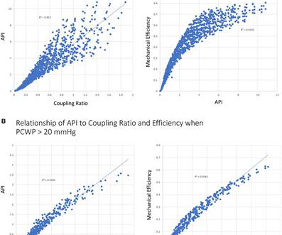
Methods Here, a mathematical and physiological framework to define the patient-specific tipping point of myocardial energetics is defined. A novel hemodynamic parameter known as the myocardial performance score (MPS), a marker of power and efficiency, is introduced that allows for the objective assessment of the physiological tipping point.
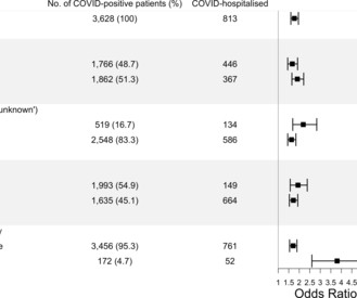
The excess risk of COVID-19 hospitalisation and death rose with increasing physiological severity of CHD (presence of pulmonary vascular disease and/or cyanosis), rather than anatomical complexity. Of patients with a positive COVID-19 test, patients with CHD were more likely than controls to be hospitalised (22.4%
Smith's ECG Blog — the presence of an almost “null vector” in standard lead I ( ie, P wave, QRS complex and T wave all under 2mm in size ) — is highly suggestive of longstanding and severe pulmonary disease. It simply does not make physiologic sense to suddenly see an all-negative QRS complex in this most lateral chest lead.
The morphology of V2-V4 is very specific in my experience for acute right heart strain (which has many potential etiologies, but none more common and important in EM than acute pulmonary embolism). CT angiogram showed extensive saddle pulmonary embolism. He had multiple cardiac arrests with ROSC regained each time. This is a quiz.
Background:Sex differences in physiology and presentation have been well established across the heart failure (HF) spectrum. Circulation, Volume 150, Issue Suppl_1 , Page A4146478-A4146478, November 12, 2024. Women had a worse Minnesota Living with Heart Failure physical subscore (34.6±5 p=0.008) and more physical signs of congestion (10.7±2.3
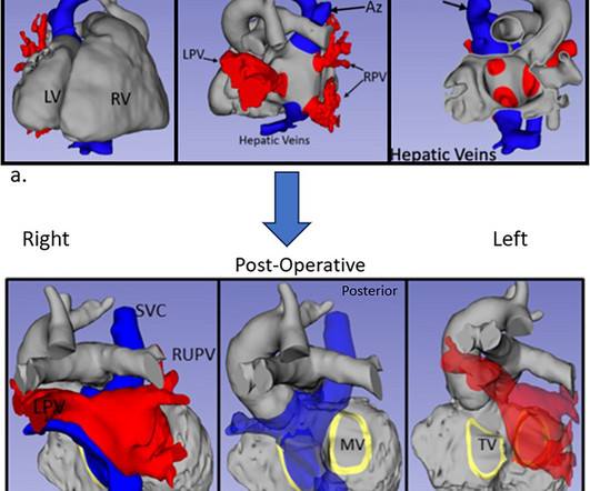
Intra-atrial baffles of pulmonary and systemic venous flows are relatively rarely used but are important surgical procedures for late-presenting d-transposition of the great arteries (D-TGA) or heterotaxy with anomalous venous connections.
This study aims to evaluate an application-based remote IHM program for infants with shunt- or duct-dependent pulmonary circulation. ConclusionApplication-based remote IHM for infants with duct- or shunt-dependent pulmonary perfusion is feasible, with high acceptance and adherence.
Of the 67 patients who underwent targeted tests, suspected diagnoses were confirmed in 49 (73%) patients: aortic stenosis (n = 8, 1%), pulmonary embolism (n = 8, 1%), seizures/stroke (n = 30, 5%), and other diseases (n = 3). Fourth, syncope in the elderly often results from polypharmacy and abnormal physiologic responses to daily events.
For right or wrong reasons, the world of electrophysiology has pushed us into a belief system that, if it is AF, the culprit must be pulmonary veins. In fact, non-pulmonary vein origins can be a staggering 70% in some series. Only 20% of focal AT arise from pulmonary veins. I guess, the same should be true for AF. Aronson JK.
Hemodynanmic of normal delivery Natural delivery involves the physiological stress of labor, which includes increased cardiac output, blood pressure fluctuations, and oxygen demand, peaking at 50-80% above baseline during contractions and pushing. In women with significant heart disease, the physiological demands of labor (e.g.,
We organize all of the trending information in your field so you don't have to. Join thousands of users and stay up to date on the latest articles your peers are reading.
You know about us, now we want to get to know you!
Let's personalize your content
Let's get even more personalized
We recognize your account from another site in our network, please click 'Send Email' below to continue with verifying your account and setting a password.


Let's personalize your content