This site uses cookies to improve your experience. To help us insure we adhere to various privacy regulations, please select your country/region of residence. If you do not select a country, we will assume you are from the United States. Select your Cookie Settings or view our Privacy Policy and Terms of Use.
Cookie Settings
Cookies and similar technologies are used on this website for proper function of the website, for tracking performance analytics and for marketing purposes. We and some of our third-party providers may use cookie data for various purposes. Please review the cookie settings below and choose your preference.
Used for the proper function of the website
Used for monitoring website traffic and interactions
Cookie Settings
Cookies and similar technologies are used on this website for proper function of the website, for tracking performance analytics and for marketing purposes. We and some of our third-party providers may use cookie data for various purposes. Please review the cookie settings below and choose your preference.
Strictly Necessary: Used for the proper function of the website
Performance/Analytics: Used for monitoring website traffic and interactions
Specific cardiovascular diseases, such as acute myocardial infarction, arrhythmias, pulmonary hypertension and pericarditis, were also pointed. IntroductionThis paper aims to expose the link between occupational exposure to respirable crystalline silica (SiO2) and cardiovascular diseases (CVDs).MethodsA
mm has been described in normal subjects) Overall impression: In my opinion and experience, this ECG most likely represents a normal baseline ECG, but with a small chance of pericarditis instead. I texted this to Dr. Smith without any information, and this was his reply: "This could be pericarditis but probably is normal variant."
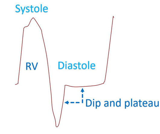
Dip and plateau pattern of ventricular pressure tracing in constrictive pericarditis Thickened and stiff pericardium in chronic constrictive pericarditis has poor compliance reducing the distension of cardiac chambers to a limited fixed total volume. Invasive hemodynamics of constrictive pericarditis. Indian Heart J.
Here is his initial ED ECG: The R-wave in V4 extends to 33 mm, the computerized QTc is 372 ms The only available previous ECG is from one year ago, during the admission when he was diagnosed with pericarditis: 1 year ago ECG, with clinician and computer interpretatioin of pericarditis Normal 0 false false false EN-US X-NONE X-NONE What do you think?
He was started on a heparin drip and CTA of the chest was ordered to rule out pulmonary embolism. This is a case like many others posted (see list below) and the EKG from the patient’s original presentation can be quickly recognized as diagnostic for pulmonary embolism. In fact, Kosuge et al. Accessed May 28, 2024. This is a quiz.
We have seen this pattern in many pts with acute right heart strain on this blog. __ Smith : The combination of T-wave inversion in V1-V3 and in lead III is very specific for acute pulmonary embolism. Acute pulmonary embolism was confirmed on CT: The patient did well with treatment. So everything about this ECG screams acute PE.
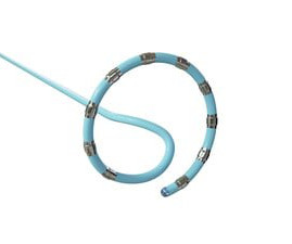
The VARIPULSE Platform is designed to enable pulmonary vein isolation with the versatility of a catheter loop, a simple generator user interface, and a mapping system that provides an intuitive, reproducible workflow with real-time visualization, contact indicator, and PF tagging mechanisms.
PR depression, which suggests pericarditis 4. We also showed that, of 47 cases of pericarditis with ST elevation, none had ST depression in aVL. ) I’ll add the following 2 comments: i ) This patient presumably has effusive-constrictive pericarditis. Absence of any ST depression in aVL. ( Clin Cardiol 22:334-344, 1999 ).
When there is MI extending all the way to the epicardium (transmural), that infarcted epicardium is often inflamed (postinfarction regional pericarditis, or PIRP). What complication is the patient with post-infarction regional pericarditis at risk for? If detected early by ultrasound, the patient can be saved. 3) Oliva et al. (4)
When there is MI extending all the way to the epicardium (transmural), that infarcted epicardium is often inflamed (postinfarction regional pericarditis, or PIRP). Rupture can be either free wall rupture (causing tamonade) or septal rupture, causing ventricular septal defect with left to right flow and resulting pulmonary edema and shock.
He was seen at another hospital and found to have a slightly elevated troponin, then underwent a CT pulmonary angiogram (PE) protocol which revealed a right sided pneumonia. Echo does not necessarily differentiate acute MI from pericarditis: both may have wall motion abnormalities. He was treated with Ceftriaxone and azithromycin.
Conduction and refractoriness alternans may be seen with WPW-related as well as AV Nodal-dependent reentr y tachycardias — atrial fibrillation — acute pulmonary embolus — myocardial contusion — and severe LV dysfunction. Therefore — identification of QRS alternans during a regular SVT does not prove the existence of an accessory pathway.
Patients with paroxysmal or persistent atrial fibrillation underwent pulmonary vein (PV) isolation under deep sedation or general anesthesia and returned for remapping at 90 days to evaluate chronic durability. paroxysmal, and 58.5% deep sedation) were treated. Eighty patients (98%) underwent remapping.
She had idiopathic ventricular fibrillation in 1992, treated with an EPD (Picture 1A), later replaced by a transvenous ICD.She was diagnosed with left femoral deep venous thrombosis and bilateral pulmonary embolism and started on therapeutic anticoagulation. However, patients with EPDs from the 1990s may present with delayed complications.
That occurs in right heart failure and constrictive pericarditis. Constrictive pericarditis is an important cause for Kussmaul sign or inspiratory increase in jugular venous pressure. On the other hand, the Y descent is very prominent in constrictive pericarditis, and it is known as Friedreich’s sign.
The morphology of V2-V4 is very specific in my experience for acute right heart strain (which has many potential etiologies, but none more common and important in EM than acute pulmonary embolism). CT angiogram showed extensive saddle pulmonary embolism. He had multiple cardiac arrests with ROSC regained each time. This is a quiz.
The combination of findings consistent with acute coronary occlusion in the anterior and inferior leads is likely due to a large "wraparound" LAD occlusion, should not be confused with the "diffuse" ST elevation of pericarditis, and will usually show reciprocal ST depression in aVL.
ECG of pneumopericardium and probable myocardial contusion shows typical pericarditis Male in 30's, 2 days after Motor Vehicle Collsion, complains of Chest Pain and Dyspnea Head On Motor Vehicle Collision. Q waves in association with RBBB are usually not seen in anterior leads unless there is pulmonary hypertension or anterior infarction.
Then the patient's pain then resolved spontaneously after 2 sublingual nitroglycerine and another ECG was recorded ECG 2 at 16 minutes ST ELEVATION CONSISTENT WITH INJURY, PERICARDITIS, OR EARLY REPOLARIZATION Overread same Smith : The T-waves are now MUCH smaller. The estimated pulmonary artery systolic pressure is 27 mmHg + RA pressure.
They include myocardial ischemia, acute pericarditis, pulmonary embolism, external compression due to mass over the right ventricular outflow tract region, and metabolic disorders like hyper or hypokalemia and hypercalcemia. According to a recent systematic review and meta-analysis, spontaneous type 1 ECG had 2.4%
The second most common cause of medical cardiac tamponade is acute idiopathic pericarditis. Less common etiologies include uremia, bacterial or tubercular pericarditis, chronic idiopathic pericarditis, hemorrhage, and other causes such as autoimmune diseases, radiation, myxedema, etc.
CT angiogram chest: no aortic dissection or pulmonary embolism. Serial chest xrays: progressive bilateral pulmonary edema. Pericarditis? No further troponins were measured. Echo: severely dilated LV, severely reduced EF at 10%, severe hypokinesis of LV from mid-apical segments with basal segments better preserved.
CT pulmonary angiogram was negative for pulmonary embolism. If the patient continues to have reperfusion, then we would expect progressive terminal T wave inversion then full T wave inversion over time in the future ECGs. Second troponin T resulted at 1,318 ng/L. Chest x-ray was read as normal. Heparin was started. Reocclusion!
In this ECG Cases blog we look at 10 cases of patients with chest pain, including false positive STEMI, false negative STEMI, and other causes to help hone your ECG interpretation skills in time-sensitive cases where those very ECG skills might save a life.
You do NOT see this in normal variant STE, nor in pericarditis. The computerized interpretation for this tracing was, “Sinus rhythm; Normal ECG” — and attention of acute care providers was apparently focused on attending to this patient’s pulmonary problems. There is upsloping ST elevation in III, with reciprocal ST depression in aVL.
The initial computer and final cardiology interpretation was a differential: “ST elevation, consider early repolarization, pericarditis, or injury.” But STEMI criteria ignore all this and look at ST segments in isolation. Based on STEMI criteria and unhelpful computer interpretation, the patient was rushed to the cath lab.
Smith : This is classic for pulmonary embolism (PE). Acute pulmonary embolism was confirmed on CT angiogram: The patient did well. See our other acute right heart strain / pulmonary embolism cases: A man in his 50s with shortness of breath Another deadly triage ECG missed, and the waiting patient leaves before being seen.
The exception is with postinfarction pericarditis , in which a completed transmural infarct results in inflammation of the subepicardial myocardium and STE in the distribution of the infarct, and which results in increased STE and large upright T-waves. These findings together are more commonly seen with pericarditis.
We organize all of the trending information in your field so you don't have to. Join thousands of users and stay up to date on the latest articles your peers are reading.
You know about us, now we want to get to know you!
Let's personalize your content
Let's get even more personalized
We recognize your account from another site in our network, please click 'Send Email' below to continue with verifying your account and setting a password.


Let's personalize your content