This site uses cookies to improve your experience. To help us insure we adhere to various privacy regulations, please select your country/region of residence. If you do not select a country, we will assume you are from the United States. Select your Cookie Settings or view our Privacy Policy and Terms of Use.
Cookie Settings
Cookies and similar technologies are used on this website for proper function of the website, for tracking performance analytics and for marketing purposes. We and some of our third-party providers may use cookie data for various purposes. Please review the cookie settings below and choose your preference.
Used for the proper function of the website
Used for monitoring website traffic and interactions
Cookie Settings
Cookies and similar technologies are used on this website for proper function of the website, for tracking performance analytics and for marketing purposes. We and some of our third-party providers may use cookie data for various purposes. Please review the cookie settings below and choose your preference.
Strictly Necessary: Used for the proper function of the website
Performance/Analytics: Used for monitoring website traffic and interactions
Sequence variants that protect against pericarditis have been discovered at a genomic locus encoding interleukin-1 immune cytokines. A newly approved drug treatment for pericarditis inhibits these cytokines, according to a new study.
Acute pericarditis (AP) is the second most common cardiac cause of chest pain, diagnosed when at least two of the following criteria are met: characteristic pleuritic chest pain, pericardial rub on auscultation, new typical ECG changes (such as widespread ST-elevation or PR-depression) and pericardial effusion on imaging.
The patient was discharged with a diagnosis of acute pericarditis — and treated with a full course of colchicine and ibuprofen. The ultimate discharge diagnosis was acute pericarditis. ( From the information provided — I would not make the diagnosis of acute pericarditis. Figure-1: The initial ECG in today's case.
Aim Anakinra, an anti IL-1 agent targeting IL-1 alfa and beta, is available for the treatment of recurrent pericarditis in cases with corticosteroid dependence and colchicine resistance after failure of conventional therapies. The efficacy endpoint was recurrence rate and the time to the first recurrence. females, 80.9%
Background Coronavirus disease (COVID-19)-associated acute pericarditis has recently received much attention owing to its high frequency associated with pericardial tamponade (PT), showing unfavorable prognosis. Conclusions We encountered a unique case of COVID-19-associated acute pericarditis exhibiting hemorrhagic PT.
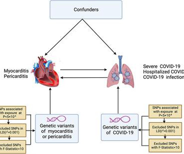
Background & aims Coronavirus disease 2019 (COVID-19) is strongly associated with myocarditis or pericarditis risk in observational studies, however, there are still studies that do not support the above conclusion. Results Non-associations in the IVW and sensitivity analyses were observed for COVID-19 with myocarditis or pericarditis.
Overall, this looks like one of the rare ECGs that is actually specific for pericarditis in my opinion. Pericarditis maybe." Meyers' words — "is one of the rare ECGs that is actually specific for pericarditis". ii ) Today's case emphasizes the importance of the history in making the diagnosis of pericarditis.
mm has been described in normal subjects) Overall impression: In my opinion and experience, this ECG most likely represents a normal baseline ECG, but with a small chance of pericarditis instead. I texted this to Dr. Smith without any information, and this was his reply: "This could be pericarditis but probably is normal variant."
"New study shows corticosteroid use may increase pericarditis recurrence in lupus patients. Researchers recommend caution with oral prednisone treatment to lowe
(MedPage Today) -- Only myocarditis/pericarditis and seizures occurred at higher rates in adolescents and children vaccinated for COVID-19 when compared with historical rates of those outcomes, according to an analysis of safety data from the FDA.
This ECG together with these symptoms is certainly concerning for OMI, but the ECG is not fully diagnostic, and another consideration could be acute pericarditis. Mistaking OMI for pericarditis is a much more harmful error than the converse. The rate is tachycardic, which is uncommon in OMI and common in pericarditis.
Why should pericarditis be considered a diagnosis of exclusion? Which clinical features are most useful in the diagnosis of pericarditis? What are the best ways to differentiate the ECG of pericarditis from that of MI and early repolarization? Why is it so important to include colchicine as part of the treatment of pericarditis?
Echocardiography-based deep learning model to differentiate constrictive pericarditis and restrictive cardiomyopathy. Commentary based on Chao CJ, Jeong J, Arsanjani R, et al. JACC Cardiovasc Imaging
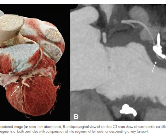
Inflammatory pericarditis can occur in differential fashion. For example, the most common chronic pericarditis tuberculosis affects the fibrinous layer. Post MI pericarditis involves the epicardium. Diastolic Coronary Artery Compression in Constrictive Pericarditis. Angina caused by calcific constrictive pericarditis.
Jesse McLaren guides us through 9 cases and explains how pericarditis is a diagnosis of exclusion through 3 simple steps: 1. Exclude complications of pericarditis, eg myocarditis, large pericardial effusion 3. The post ECG Cases 27 Pericarditis – Diagnosis of Exclusion appeared first on Emergency Medicine Cases.
The computer interpretation was “ST elevation, consider early repolarization, pericarditis or injury.” The final cardiology interpretation confirmed the computer interpretation of “ST elevation, consider early repolarization, pericarditis or injury”. A healthy 45-year-old female presented with chest pain, with normal vitals.
BackgroundAcute pericarditis represents an inflammatory disease affecting the pericardial layers. Case presentationWe reported a case of a patient with acute pericarditis that occurred a few hours after chest radiation therapy, performed for breast cancer.
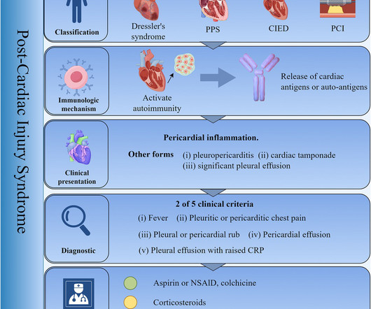
Post-Cardiac Injury Syndrome (PCIS) refers to a collective term encompassing post-myocardial infarction syndrome, post-pericardiotomy syndrome, and post-traumatic pericarditis. This condition is considered to be an autoimmune-mediated inflammatory response leading to pericarditis as the primary manifestation of the cardiac disease.
Clinician and EKG machine read of acute pericarditis. While it is true that inferior MI has ST depression in aVL 99% of the time (Bischof and Smith), and that inferolateral ST elevation is the most common distribution for pericarditis, the ST elevation in V3 has "terminal QRS distortion (TQRSD)," (diagnostic of LAD occlusion).
DALLAS, June 17, 2024 — About 40,000 people in the United States experience recurrent pericarditis, or inflammation of the sac-like structure that protects the heart, which can cause chest pain and may lead to fluid buildup around the heart muscle.
But there was some doubt as to whether it might be pericarditis because of the ST elevation in I and II, without ST depression in III. Add that to "sharp" pain and a 33 year old, and it is easy to convince yourself that this is, indeed, pericarditis. This is a good sign for myocardial infarction and does not happen in pericarditis.
Pericarditis refers to inflammation of the pericardium The pericardium is a sac within which the heart sits. Acute inflammation of this sac is known as acute pericarditis. About 5% of patients who present to A+E with chest pain which is not deemed to be a heart attack or angina are ultimately diagnosed with pericarditis.
Background There are limited data on acute pericarditis according to different age groups. The aim of this study is to investigate the role of age-related features in clinical characteristics, management, and outcomes of acute pericarditis, with a focus on the geriatric population. and G4: 16.2%; p<0.001). to 5.58, p<0.001).
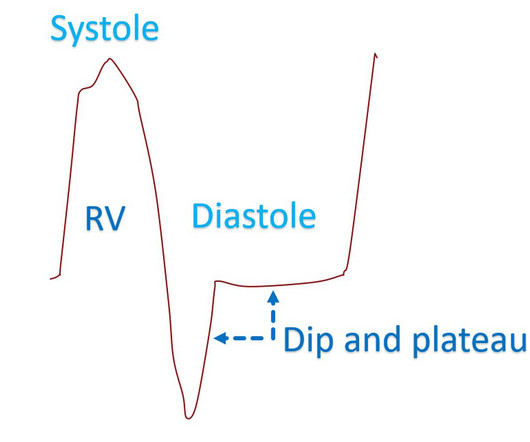
Dip and plateau pattern of ventricular pressure tracing in constrictive pericarditis Thickened and stiff pericardium in chronic constrictive pericarditis has poor compliance reducing the distension of cardiac chambers to a limited fixed total volume. Invasive hemodynamics of constrictive pericarditis. Indian Heart J.
Here is his initial ED ECG: The R-wave in V4 extends to 33 mm, the computerized QTc is 372 ms The only available previous ECG is from one year ago, during the admission when he was diagnosed with pericarditis: 1 year ago ECG, with clinician and computer interpretatioin of pericarditis Normal 0 false false false EN-US X-NONE X-NONE What do you think?
Transition to rilonacept monotherapy from oral therapies in patients with recurrent pericarditis. Brucato A, Wheeler A, Luis SA, et al. Heart 2023; 109 : 297-304. This article has been corrected since it was first published.
Specific cardiovascular diseases, such as acute myocardial infarction, arrhythmias, pulmonary hypertension and pericarditis, were also pointed. IntroductionThis paper aims to expose the link between occupational exposure to respirable crystalline silica (SiO2) and cardiovascular diseases (CVDs).MethodsA
TB pericarditis is the commonest cardiac manifestation of TB and is the leading cause of constrictive pericarditis, a reversible (by surgical pericardiectomy) cause of diastolic heart failure in endemic areas.
To investigate the relationship between p wave terminal force (Ptfv1) and pericardial thickness in patients with tuberculous constrictive pericarditis.
06:44 - T-waves in V2 are smaller now - Overall resolution of prior findings (which qualifies as a dynamic change) The initial note by the cardiologist states that the presentation is more consistent with pericarditis. Remember, pericarditis is the thing you say and write down when youre actively trying to miss an OMI.
Acute pericarditis is characterized by pericardial inflammation which can be treated with anti-inflammatory drugs. A considerable percentage of patients develops recurrent pericarditis with several relapses. Two pathophysiological mechanisms have been described for idiopathic recurrent pericarditis, autoimmune and autoinflammatory.
Jesse McLaren guides us through 10 cases, driving home the points that sepsis is a common cause of rapid Afib and diffuse ST depression with reciprocal ST elevation in aVR, myo/pericarditis is a diagnosis of exclusion, endocarditis or lyme carditis can cause AV block, PE can cause low grade fever and ECG signs of acute RV strain and that fever can (..)
A new study of more than 2,900 patients provides evidence that it's likely best to use as little corticosteroid medicine as possible when treating people who have lupus pericarditis, a common heart complication of the autoimmune disease Systemic Lupus Erythematosus (SLE).
Pericarditis is the most common complication following hybrid sinus node sparing ablation for Inappropriate Sinus Tachycardia (IST)/Postural Orthostatic Tachycardia Syndrome (POTS).
Recurrent pericarditis (RP) is the most troublesome complication of acute pericarditis reflecting an unresolving inflammation of the pericardial sac around the heart and associated with significant morbidity.
This is a value typical for a large subacute MI, n ormal value 48 hours after myocardial infarction is associated with Post-Infarction Regional Pericarditis ( PIRP ). As already mentioned, this patient could have post-infarction regional pericarditis from a large completed MI. Sinus tachycardia has many potential causes. Hammill SC.
A 69-year-old woman with a history of focal pleural plaques due to occupational asbestos exposure presented with chronic dyspnea on exertion. Chest imaging showed calcification of the pericardium.
Publication date: Available online 20 May 2024 Source: The American Journal of Cardiology Author(s): Shaye Kivity, Tomer Ziv Baran, Miri Mizrahi Reuveni, Angela Irony, Limor Adler, Yehuda Alder, Roma Parikh, Sara Kivity
The undergraduate continues: This new EKG pattern is more suggestive of acute pericarditis. Usually with pericarditis, some degree of PR segment depression is expected. This is typical of pericarditis. But, as I always say, you diagnose pericarditis at your peril. This EKG seems to lack it.
It is easy to say pericarditis in such a case. young male no risk factors and ST-elevation in several leads) As Dr. Smith has emphasized many times you diagnose pericarditis at your patient's and your own peril. Version 1 was not trained to detect myo- or pericarditis. The above ECG was recorded. How did the Queen do?
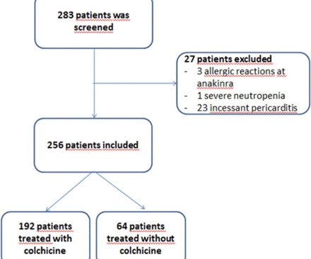
Objective Clinical trials have evaluated the efficacy and safety of colchicine only in simple pericarditis, excluding cases of concomitant myocarditis. The aim of this paper is to evaluate the efficacy and safety of colchicine for the treatment of the first attack of acute pericarditis with concomitant myocardial involvement.
We organize all of the trending information in your field so you don't have to. Join thousands of users and stay up to date on the latest articles your peers are reading.
You know about us, now we want to get to know you!
Let's personalize your content
Let's get even more personalized
We recognize your account from another site in our network, please click 'Send Email' below to continue with verifying your account and setting a password.


Let's personalize your content