This site uses cookies to improve your experience. To help us insure we adhere to various privacy regulations, please select your country/region of residence. If you do not select a country, we will assume you are from the United States. Select your Cookie Settings or view our Privacy Policy and Terms of Use.
Cookie Settings
Cookies and similar technologies are used on this website for proper function of the website, for tracking performance analytics and for marketing purposes. We and some of our third-party providers may use cookie data for various purposes. Please review the cookie settings below and choose your preference.
Used for the proper function of the website
Used for monitoring website traffic and interactions
Cookie Settings
Cookies and similar technologies are used on this website for proper function of the website, for tracking performance analytics and for marketing purposes. We and some of our third-party providers may use cookie data for various purposes. Please review the cookie settings below and choose your preference.
Strictly Necessary: Used for the proper function of the website
Performance/Analytics: Used for monitoring website traffic and interactions
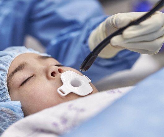
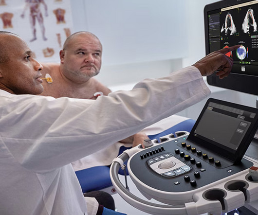
Cardiovascular ultrasound has played a key role in the evolution of early diagnosis of structural heart disease, led by a technology pioneered by Philips: the ‘transesophageal echocardiography’ (TEE) ultrasound transducer. TEE helps cardiologists by providing highly detailed images of the heart and its internal structures.
Image courtesy: Philips christine.book Wed, 06/12/2024 - 14:07 June 12, 2024 — Royal Philips has announced its next-generation AI-enabled cardiovascular ultrasound platform to help speed up cardiac ultrasound analysis with proven AI technology and reduce the burden on echocardiography labs.
Zane was diagnosed during a routine ultrasound at 20 weeks’ gestation and his mom, Kayla, was immediately referred to the Colorado Fetal Care Center (CFCC) at Childrens Hospital Colorado. Consistently ranked in the top 10 pediatric cardiology programs by U.S.
Getty Images milla1cf Tue, 12/12/2023 - 09:04 December 12, 2023 — The American Society of Echocardiography (ASE) and the ASE Foundation have awarded grant funding totaling $75,000 for three innovative cardiovascular ultrasound research projects led by early career investigators. Three recipients were each awarded a $25,000 grant.
Ultrasound techniques currently used in echocardiography uses frame rates from 30-150 frames/s. This limits its temporal resolution for very short lived events, especially in pediatric and congenital heart disease with faster heart rates compared to adults [1]. J Am Coll Cardiol. 2024 Jan 2;83(1):63-81. doi: 10.1016/j.jacc.2023.10.025.
This year’s program, “Global Echocardiography: Innovations in Diagnosis and Beyond,” is described as an educational experience gathering global experts and enthusiasts in echocardiography, hosted by the largest global organization for cardiovascular ultrasound imaging serving physicians, sonographers, nurses, and scientists.
Background Axillary arterial access (AAA) in pediatric heart catheterizations is undervalued. We aimed ultrasound-guided punctures in the proximal two-thirds of axillary arteries with diameters ≥2 mm to insert 7 cm/4 Fr short introducers.
Developed at Children’s National Hospital and detailed in the latest edition of the Journal of the American Heart Association , the new AI system combines the power of novel ultrasound probes with portable electronic devices installed with algorithms capable of diagnosing RHD on echocardiogram. Beginning in March, Craig Sable, M.D.
Fetal cardiac intervention team included pediatric cardiology imaging specialist, fetal cardiology nurse practitioner, two interventional pediatric cardiologists, ultrasound radiologist, maternal-fetal medicine physician, fetal anaesthesiologist and maternal anaesthesiologist. Reference Ryan Callahan, Kevin G. Esch, Lynn A.
Methods We performed a retrospective analysis to examine the clinical manifestations, genetic traits, and the relationship between PD and mitochondrial function in a pediatric patient.
For example, by integrating Ventripoint’s AI-powered heart-scanning technology, which turns ultrasound images of the heart into MRI-quality heart images, InView provides pediatric cardiologists with access to MRI-quality heart images at a fraction of the cost and time needed for traditional MRIs. As well, by incorporating Us2.ai’s
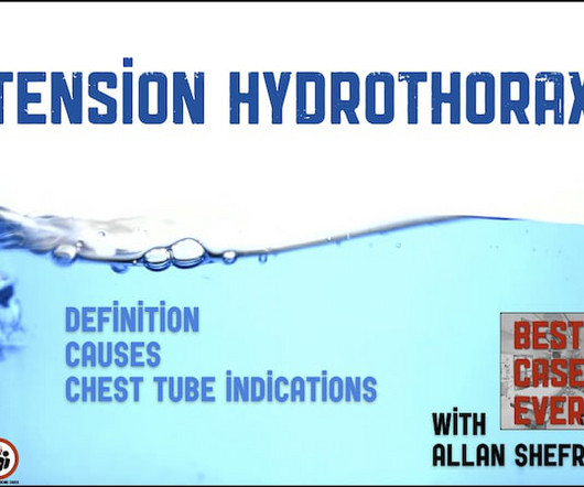
Allan Shefrin tells his Best Case Ever of a child who presents in shock and discusses the causes of tension hydrothorax, indications for tube thoracostomy for hydrothorax and integration of POCUS into pediatric resuscitation. The post BCE 81 Tension Hydrothorax appeared first on Emergency Medicine Cases.
And Regulatory Issues regarding medical devices and drug development — Rob Kazmierski (FDA) A Center for Drug Perspective on Cardiac Safety — Rosilyn Adigun (FDA) Consumer Wearables in pediatrics: Should there be standards?
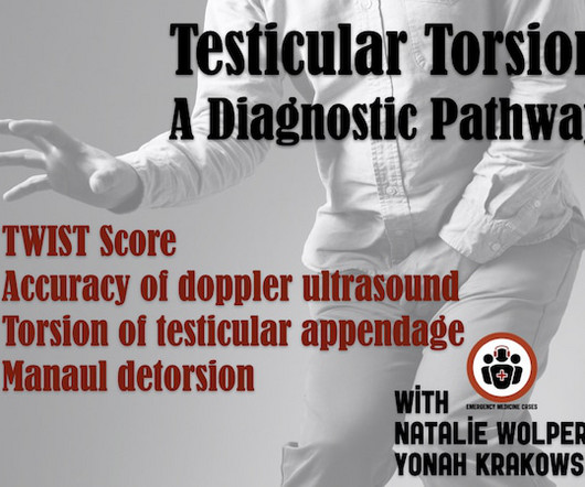
How accurate is doppler ultrasound in the diagnosis of testicular torsion? Are there any set of clinical symptoms and signs or decision tools (such as the TWIST Score) that can rule in or rule out testicular torsion with confidence? To what degree does Prehn's sign help distinguish epididymitis from testicular torsion?
Hemodynamic instability in trauma is usually due to bleeding, but if ultrasound shows poor contractility, then this may be due to cardiac contusion. In the ED, ultrasound showed hemopericardium with tamponade. Outcome Three weeks later, shortly after having been physically active (bouncing on a trampoline), she was found unresponsive.
Food and Drug Adminstration (FDA) has approved DEFINITY (Perflutren Lipid Microsphere) as an ultrasound enhancing agent for use in pediatric patients with suboptimal echocardiograms, including those who have undergone heart transplant, or have Kawasaki disease or a congenital cardiovascular anomaly. Lantheus announced that the U.S.
A bedside cardiac ultrasound was normal. This case was sent by Dr Avinash Krishnamurthy, a fine emergency medicine resident from Australia Cairns base hospital Case : An adolescent male had a mechanical fall and injured his left shoulder and arm. His chest was tender. An ECG was recorded: Avinash was understandably confused by this ECG.
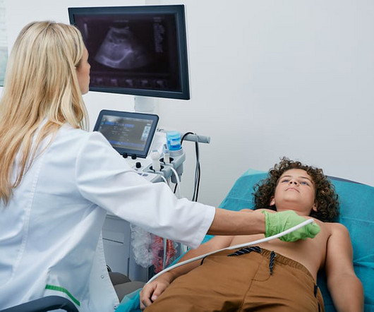
During echocardiography, a transducer transmits the ultrasound beam towards the heart. Subcostal view is a favourite view of pediatric echocardiographers. It is used in the emergency department, at bedside, in the intensive care unit as well as in the operating room. Hence a basic knowledge is needed for all physicians and paramedics.
To create comprehensive reports, connect to applications dealing with 4D, echocardiography, nuclear medicine, CT angiography, and pediatric echo reporting. This is a great way to increase productivity in any medical setting.
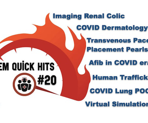
Justin Morgenstern on imaging choices in renal colic, Hanni Stoklosa on recognition and management of human trafficking, Rohit Mohindra on management of atrial fibrillation during COVID-19, Anand Swaminathan on transvenous pacemaker placement, Rob Simard on COVID-19 lung POCUS, Brit Long on COVID-19 dermatology and Sarah Foohey & Paul Koblic on (..)
A bedside cardiac ultrasound revealed grossly normal to hyperdynamic systolic function with no obvious areas of wall motion abnormalities. This was recorded about 30 minutes later: Same A previous ECG was obtained and was normal. The patient denied any chest pain whatsoever, and a troponin at zero and 2 hours were both undetectable.
We organize all of the trending information in your field so you don't have to. Join thousands of users and stay up to date on the latest articles your peers are reading.
You know about us, now we want to get to know you!
Let's personalize your content
Let's get even more personalized
We recognize your account from another site in our network, please click 'Send Email' below to continue with verifying your account and setting a password.


Let's personalize your content