This site uses cookies to improve your experience. To help us insure we adhere to various privacy regulations, please select your country/region of residence. If you do not select a country, we will assume you are from the United States. Select your Cookie Settings or view our Privacy Policy and Terms of Use.
Cookie Settings
Cookies and similar technologies are used on this website for proper function of the website, for tracking performance analytics and for marketing purposes. We and some of our third-party providers may use cookie data for various purposes. Please review the cookie settings below and choose your preference.
Used for the proper function of the website
Used for monitoring website traffic and interactions
Cookie Settings
Cookies and similar technologies are used on this website for proper function of the website, for tracking performance analytics and for marketing purposes. We and some of our third-party providers may use cookie data for various purposes. Please review the cookie settings below and choose your preference.
Strictly Necessary: Used for the proper function of the website
Performance/Analytics: Used for monitoring website traffic and interactions
Bedside ED ultrasound showed exceedingly poor global LV function, and no B lines. Pacemaker mediated tachycardia! Pacemaker mediated tachycardia , also called "Endless Loop Tachycardia," cannot happen during atrial fibrillation, so the A fib must have converted. Here is the initial ED ECG. What do you think?
milla1cf Thu, 05/02/2024 - 10:09 May 2, 2024 — Artificial intelligence experts at Cedars-Sinai and the Smidt Heart Institute created a dataset with more than 1 million echocardiograms, or cardiac ultrasound videos, and their corresponding clinical interpretations. Image by Getty.
The post EM Quick Hits 20 Imaging Renal Colic, Human Trafficking, Atrial Fibrillation During COVID, Transvenous Pacemaker Placement, COVID Lung POCUS, COVID Derm, Virtual Simulation appeared first on Emergency Medicine Cases.
Bedside cardiac ultrasound showed moderately decreased LV function. Place temporary pacemaker 3. (And of course Ken's comments at the bottom) An elderly obese woman with cardiomyopathy, Left bundle branch block, and chronic hypercapnea presented hypoxic with altered mental status. She was intubated.
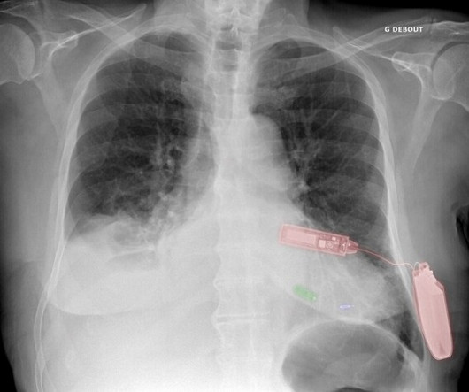
Green: Micra leadless pacemaker; blue: WiSE-CRT system LV endocardial electrode; and red: WiSE-CRT system subcutaneous battery and ultrasound generator. Carabelli A, Jabeur M, Jacon P, Rinaldi CA, European experience with a first totally leadless cardiac resynchronization therapy pacemaker system. 2021 May 21;23(5):740-747.
At AMS Cardiology, we offer comprehensive cardiac care including services tailored to the specific needs of seniors such as: Echocardiography: This painless ultrasound uses sound waves to create images of your heart, allowing doctors to assess its structure and function.
Automatic activity refers to enhanced pacemaking function (typically from a non sinus node source), for example atrial tachycardia. However the patient continued to have chest pain and bedside ultrasound showed hypokinesis of the septum with significantly reduced LVEF. The most common triggered arrhythmia is Torsades de Pointes.
Discussing further, Catheterization Laboratory, also called Cath Lab, is a medical examination room where angiogram, angioplasty, ablation, and implantation of pacemaker are carried out. Ultrasound, TEE, and IVUS play an influential role in dropping the usage of angiographic imaging. Building Smart Cath Labs is highly related to it.
At AMS Cardiology, we offer comprehensive cardiac care including services tailored to the specific needs of seniors such as: Echocardiography: This painless ultrasound uses sound waves to create images of your heart, allowing doctors to assess its structure and function.
A bedside cardiac ultrasound was normal. A Patient with Ischemic symptoms and a Biventricular Pacemaker This case was sent by Dr Avinash Krishnamurthy, a fine emergency medicine resident from Australia Cairns base hospital Case : An adolescent male had a mechanical fall and injured his left shoulder and arm. His chest was tender.
If there had been — a temporary atrial pacemaker could have been considered as a way of increasing the heart rate to suppress a bradycardia-dependent arrhythmia ("overdrive pacing"). Another approach is sympathetic chain (stellate ganglion) blockade if you have the skills to do it: it requires some expertise and ultrasound guidance.
Cross Compatibility The easy-to-install SoftCath software can easily be integrated and used for Angiography, Pacemaker, Valvuloplasty, ICD, etc. Reports can be prepared for ultrasound, angioplasty, and TEE with the use of robust analysis. Therefore, one can make use of one cardiology reporting software instead of multiple software.
This is an ultrasound (a bit like the type that we use on pregnant women to look at the baby). An ultrasound will allow you to visualise the heart, measure the sizes of the chambers, assess the heart valves and work out how well the heart functions as a pump. It is a super specialised discipline. It requires very expensive machinery.
Dr. Nossen performed a bedside ultrasound which was interpreted as normal. Nossen needed no AI and interpreted this as ectopic atrial rhythm (ectopic atrial escape as the heart rate is in the range of 48 bpm), with normal variant ST elevation likely exaggerated by atrial repolarization (termed "Ta" wave).
Cupid EHR from Epic boasts the following: Cloud-based EHR Offers integrated order entry, scheduling, procedure documentation, structured reporting, and data analytics for cardiology practices Supports a wide range of workflows, including Echocardiograms, Ultrasound vascular, Cardiac Cath, stress testing, Electrophysiology, and structured documentation (..)
Her bedside cardiac ultrasound was normal We decided to cardiovert her since the time of onset was very recent. But when you see this, you should suspect that the AV node is not well. Our electrophysiologist told me that highly trained athletes can have such high vagal tone that they do not have a rapid ventricular response.
Check : [vitals, SOB, Chest Pain, Ultrasound] If the patient has Abdominal Pain, Chest Pain, Dyspnea or Hypoxemia, Headache, Hypotension , then these should be considered the primary chief complaint (not syncope). Negative predictors included dementia, pacemaker, coronary revascularization, and cerebrovascular disease.
If you do not have an arterial line, use bedside ultrasound to verify myocardial contractility corresponds to pacing. If you don't have ultrasound (but you should), then palpate a pulse! Electrical artifact is usually "blocked out" from the ECG monitor ( by integrated pacemaker software that eliminates a 40-to-80 msec.
We organize all of the trending information in your field so you don't have to. Join thousands of users and stay up to date on the latest articles your peers are reading.
You know about us, now we want to get to know you!
Let's personalize your content
Let's get even more personalized
We recognize your account from another site in our network, please click 'Send Email' below to continue with verifying your account and setting a password.


Let's personalize your content