This site uses cookies to improve your experience. To help us insure we adhere to various privacy regulations, please select your country/region of residence. If you do not select a country, we will assume you are from the United States. Select your Cookie Settings or view our Privacy Policy and Terms of Use.
Cookie Settings
Cookies and similar technologies are used on this website for proper function of the website, for tracking performance analytics and for marketing purposes. We and some of our third-party providers may use cookie data for various purposes. Please review the cookie settings below and choose your preference.
Used for the proper function of the website
Used for monitoring website traffic and interactions
Cookie Settings
Cookies and similar technologies are used on this website for proper function of the website, for tracking performance analytics and for marketing purposes. We and some of our third-party providers may use cookie data for various purposes. Please review the cookie settings below and choose your preference.
Strictly Necessary: Used for the proper function of the website
Performance/Analytics: Used for monitoring website traffic and interactions
Pacemaker mediated tachycardia! Pacemaker mediated tachycardia , also called "Endless Loop Tachycardia," cannot happen during atrial fibrillation, so the A fib must have converted. Another ECG was recorded 12 minutes later: Paced rhythm, probable Pacemaker-Mediated Tachycardia ? The patient was admitted without angiogram."
Cardiac pacemaker device implantation is associated with incidence of different complications. We report a case of combined chylothorax and pulmonary embolism following a dual chamber pacemaker implantation.
Introduction Multiple abnormal electrocardiographic findings have been documented in patients experiencing acute pulmonary embolism. To date, only a limited number of cases involving a complete atrioventricular block have been reported in acute pulmonary embolism. Echocardiography confirmed signs of right ventricular dysfunction.
The pulmonary vein (PV) sleeves are a major source of ectopic activity driving atrial fibrillation (AF) and episodes of AF in patients are more prevalent at night.
We found in our last trial, REDUCE LAP-HF II, that HFpEF patients with pacemakers or pulmonary vascular disease didn't benefit from atrial shunting. In our ongoing RESPONDER-HF trial we are excluding those patients and using exercise hemodynamics to qualify and randomize HFpEF patients most likely to respond favorably to shunting."
Due to atrial and ventricular pacing dependence, a comprehensive congenital care team concluded the need for lead extraction and replacement of pacemaker via leadless peacemaking device. Laser-lead extraction and temporary atrial pacemaker placement was performed.
This is demonstrated ( Figure 5 ) by the gap in arrows at the bottom of the strip, signifying that the demand pacemaker has recognized an underlying rhythm (in this case, artifact from a moving ambulance). The artifact fools the pacemaker into thinking the rhythm is native. On ED arrival ROSC is achieved.
Unfortunately, patients will often present late in their disease course with severe right-sided heart failure, pulmonary hypertension, and life-limiting symptoms that have few durable treatment options. Tricuspid valve disease is an often underrecognized clinical problem that is associated with significant morbidity and mortality.
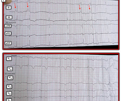
This middle-aged patient presented with SOB, weakness, and mild pulmonary edema. She had a permanent pacemaker implanted. After pacer AND conversion to sinus rhythm: Computer diagnosis: IMPRESSION ELECTRONIC VENTRICULAR PACEMAKER ABNORMAL RHYTHM ECG What is missing from this interpretation? This shows atrial fibrillation.
These 2 settings are: i ) In patients with severe , often longstanding pulmonary disease ; and / or , ii ) In acutely ill patients with multi-system disease ( ie, sepsis, shock, electrolyte and/or acid-base disorders ). MAT is not a Wandering Pacemaker. Hypoxic injury ( from pneumonia or other acute pulmonary complication ).
Recently, improvements in the repair of tetralogy of Fallot have increased the need for reoperation in adulthood, and it’s not rare that these reoperation candidates suffer from biventricular failure. However,
CT of the chest showed no pulmonary embolism but bibasilar infiltrates. Place temporary pacemaker 3. (And of course Ken's comments at the bottom) An elderly obese woman with cardiomyopathy, Left bundle branch block, and chronic hypercapnea presented hypoxic with altered mental status. She was intubated.
Tricuspid valve replacement was performed in 26 (53%) patients including 19 (73%) cases of combined pulmonary valve replacement. Epicardial pacemakers were systematically implanted in operated patients and 25% were permanently paced. A postoperative positive right ventricular remodelling was observed (p<0.001).
Patients with more severe obesity were more likely to have responder characteristics for atrial shunt therapy (fewer pacemakers and lower exercise pulmonary vascular resistance [PVR]). Pulmonary vascular resistance at rest and exercise decreased with higher BMI.
T wave alternans is characterized by variation in T-wave morphology in the setting of consistent pacemaker and QRS morphology. (1) She had an uneventful ICU course and was extubated for ongoing care with the inpatient psychiatric service. Teaching Points: 1. NOTE : On occasion — Alternans may be seen with monomorphic VT ( Maury and Metzger ).
I’d guess the overall rhythm is sinus, perhaps with a wandering atrial pacemaker and very frequent ventricular ectopy with multiple couplets. Perhaps the patient has pulmonary hypertension and/or tricuspid regurgitation? In the meantime, a pacemaker may be needed. =
Oral anticoagulation also reduced a composite of cardiovascular death, all-cause stroke, peripheral arterial embolism, myocardial infarction or pulmonary embolism (RR 0.85, 95% CI 0.73-1.00, We used random-effects models for meta-analysis and rated the quality of evidence using the GRADE framework. 1.00, I2=0%; moderate-quality evidence).
Recursive feature elimination was employed to identify the most relevant features in predicting the risk of mortality. Abstract Aims Accurate selection of patients with severe heart failure (HF) who might benefit from advanced therapies is crucial.
ECG Blog #65 — for an example of MAT in a patient with chronic pulmonary disease ( plus more on the differential diagnosis of MAT ). ECG Blog #200 — for an example of Wandering Atrial Pacemaker. CASE Follow-Up: Lab work that had been drawn before the cardiac arrest — came back after the arrest.
It is also not a wandering pacemaker — because change in atrial pacing site is gradual with that disorder. Having observed this phenomenon over many years — I’ve noticed that rather than black-or-white classifications for rhythms such as wandering pacemaker; sinus with many PACs; and MAT — that there is a s pectrum for these rhythm disorders.
Thrombus can sometimes occur when there is a central venous catheter or a multiple pacemaker or defibrillator leads there that can cause thrombus formation. Right atrial hypertrophy as in tricuspid stenosis, pulmonary stenosis and pulmonary hypertension. But in a VSD with pulmonary hypertension A wave is not prominent.
MAT has at least 3 distinct P-wave morphologies, but there is no single dominant pacemaker (i.e., Given the significant tachycardia and this “middle-ground” irregular SVT rhythm ( regardless of what you call it ) — acute pulmonary disease is virtually certain to be an important contributing ( if not the sole ) cause of this tachyarrhythmia.
Methods Octogenarians with AF or consecutive atrial tachycardia undergoing index or re-ablation (pulmonary vein isolation [PVI] and ablation beyond PVI with different energy sources) in a single center, were analyzed. However, concomitant infections and pacemaker implantations occur in this cohort. Hospital stay after CA was 2.32
We assess dLVEDPs accuracy in predicting pulmonary capillary wedge pressure (PCWP) and propose a corrective equation.METHODS:We included 29 consecutive patients treated with Impella 5.5: performance level, heart rhythm, pacemaker settings, sex, mechanical ventilation, and body mass index were recorded. Variables such as Impella 5.5
Negative predictors of adverse outcome: Pacemaker Pre-syncope or "near-syncope," but there is still some small risk (5, 18) These last two are identified in studies, but I consider them dangerous signs and symptoms in their own right, as above: 10. QRS Aortic Dissection, Valvular (especially Aortic Stenosis), Tamponade. of ED visits.
These issues can only be addressed in an ICCU (Intensive Coronary Care Unit) setting, where temporary pacemakers and defibrillators are available. Therefore, seeking early admission to the ICCU is the initial step toward addressing such problems.
We organize all of the trending information in your field so you don't have to. Join thousands of users and stay up to date on the latest articles your peers are reading.
You know about us, now we want to get to know you!
Let's personalize your content
Let's get even more personalized
We recognize your account from another site in our network, please click 'Send Email' below to continue with verifying your account and setting a password.


Let's personalize your content