This site uses cookies to improve your experience. To help us insure we adhere to various privacy regulations, please select your country/region of residence. If you do not select a country, we will assume you are from the United States. Select your Cookie Settings or view our Privacy Policy and Terms of Use.
Cookie Settings
Cookies and similar technologies are used on this website for proper function of the website, for tracking performance analytics and for marketing purposes. We and some of our third-party providers may use cookie data for various purposes. Please review the cookie settings below and choose your preference.
Used for the proper function of the website
Used for monitoring website traffic and interactions
Cookie Settings
Cookies and similar technologies are used on this website for proper function of the website, for tracking performance analytics and for marketing purposes. We and some of our third-party providers may use cookie data for various purposes. Please review the cookie settings below and choose your preference.
Strictly Necessary: Used for the proper function of the website
Performance/Analytics: Used for monitoring website traffic and interactions
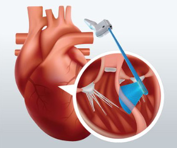
Currently, TEE cannot be leveraged during cardiac bypass surgery. Instead, a surgeon views the patient’s septal thickness via TEE prior to performing open-heartsurgery and would trim the septum – if necessary – without real-time guidance.
Zane had three openheartsurgeries before he turned 3. Zeke and Zane were both diagnosed with hypoplastic left heart syndrome (HLHS) before birth. As far as congenital heart diseases go, HLHS falls on the rarer end of the spectrum. And surprisingly, he had a more routine care journey than his brother, Zeke.
The Warrior Heart Project Hi my name is Addison Gutierrez! On June 21, 2008 my mom went in for a routine ultrasound. My dad was a surgery resident at the hospital so he was able to meet her there. It had been missed on my ultrasound. I was rushed to the cath lab for emergency surgery.
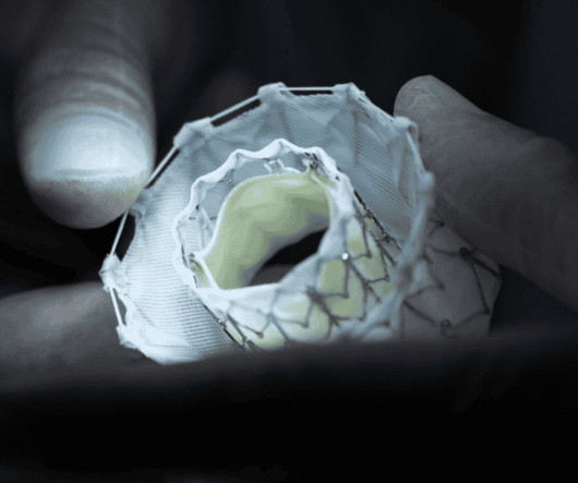
Focusing on mitral and tricuspid valve diseases , Capstans treatment combines transcatheter implantation of a folded valve replacement with its X-ray and ultrasound-guided robot to align the low-profile implant with the beating heart valve.
When we first learned that Austin would be admitted to Rocky Mountain Hospital for Children PICU for openheartsurgery at 3 weeks old, we were nervous and anxious for what was ahead. He was diagnosed with a butterfly vertebrae, kidney fullness, sacral dimple, and several heart defects (right aortic arch, VSD, and ASD).
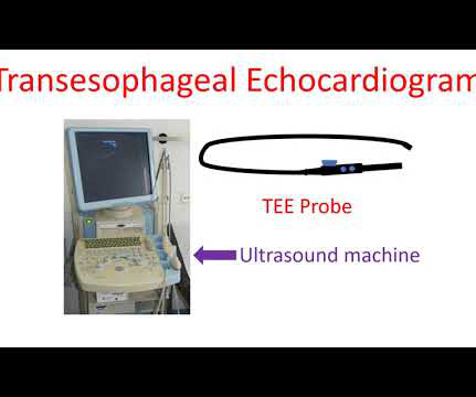
Echocardiogram is an image of the heart using ultrasound. An ultrasound beam is transmitted into the body using a device known as transducer. The echo received from the body is processed by the computer in the machine to give an image of the heart.
We organize all of the trending information in your field so you don't have to. Join thousands of users and stay up to date on the latest articles your peers are reading.
You know about us, now we want to get to know you!
Let's personalize your content
Let's get even more personalized
We recognize your account from another site in our network, please click 'Send Email' below to continue with verifying your account and setting a password.


Let's personalize your content