Remove Myocardial Infarction Remove SCAD Remove STEMI
article thumbnail

Differences in left ventricular myocardial function and infarct size in female patients with ST elevation myocardial infarction and spontaneous coronary artery dissection

Frontiers in Cardiovascular Medicine

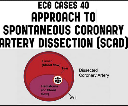
Introduction Differences in pathophysiology, clinical presentation, and natural course of ST-elevation myocardial infarction in female patients due to either spontaneous dissection (SCAD-STEMI) or atherothrombotic occlusion (type 1 STEMI) have been discussed. vs. 1.8 ± 5.1%, p  = 0.002).

article thumbnail

ECG Cases 40 – Approach to Spontaneous Coronary Artery Dissection (SCAD)

ECG Cases

Jesse McLaren on when to consider Spontaneous Coronary Artery Dissection (SCAD), which patients are at risk for reocclusion, and the challenges of diagnosing SCAD in patients who have nonischemic ECGs despite silent occlusion, occlusions perfused by collaterals, or from non-occlusive MI on this ECG Cases.

SCAD 52
article thumbnail

Management and outcomes of spontaneous coronary artery dissection: a systematic review of the literature

Frontiers in Cardiovascular Medicine

Background Contemporary management of spontaneous coronary artery dissection (SCAD) is still controversial. Results The systematic review included 13 observational studies evaluating 1,801 patients with SCAD. Results The systematic review included 13 observational studies evaluating 1,801 patients with SCAD. Approximately 48.5%

SCAD 75
article thumbnail

A young peripartum woman with Chest Pain

Dr. Smith's ECG Blog

In the absence of these factors it is termed spontaneous coronary artery dissection ( SCAD ). At that time the literature suggested: SCAD was rare , Mostly related to pregnancy , Seen on angiography as a dissection flap , and Managed similarly to MI caused by CAD (ASA, BB, lytics/PCI ). The SCAD cases in Lobo et al. Lobo et al.

article thumbnail

A woman in her 30s with sudden chest pain, nausea, and diaphoresis. Was her cardiology management appropriate?

Dr. Smith's ECG Blog

There is clearly sufficient STE for STEMI criteria in leads V2 and aVL, but lead I has less than 1.0 mm of STE - thus, technically this ECG does not meet STEMI criteria, although it is a quite obvious OMI. This ECG was immediatel y discussed with the on-call cardiologist who said the ECG was "concerning but not a STEMI."

article thumbnail

An athletic 30-something woman with acute substernal chest pressure

Dr. Smith's ECG Blog

It is equivalent to a transient STEMI. This is diagnostic of myocardial infarction. Not much, but studies of STEMI and NonSTEMI show that about 70% of those diagnosed with STEMI have a peak troponin I above 10 ng/mL and that about 70% of those diagnosed with NonSTEMI have a peak troponin I below 10 ng/mL.

SCAD 52
article thumbnail

1 hour of CPR, then ECMO circulation, then successful defibrillation.

Dr. Smith's ECG Blog

This is a troponin I level that is almost exclusively seen in STEMI. In this case, profound shock for 1 hour would result in the same degree of infarction. A followup ECG was recorded 2 days later: No definite evidence of infarction. So this is either a case of MINOCA, or a case of Type II STEMI. Troponin I rose to 44.1