This site uses cookies to improve your experience. To help us insure we adhere to various privacy regulations, please select your country/region of residence. If you do not select a country, we will assume you are from the United States. Select your Cookie Settings or view our Privacy Policy and Terms of Use.
Cookie Settings
Cookies and similar technologies are used on this website for proper function of the website, for tracking performance analytics and for marketing purposes. We and some of our third-party providers may use cookie data for various purposes. Please review the cookie settings below and choose your preference.
Used for the proper function of the website
Used for monitoring website traffic and interactions
Cookie Settings
Cookies and similar technologies are used on this website for proper function of the website, for tracking performance analytics and for marketing purposes. We and some of our third-party providers may use cookie data for various purposes. Please review the cookie settings below and choose your preference.
Strictly Necessary: Used for the proper function of the website
Performance/Analytics: Used for monitoring website traffic and interactions
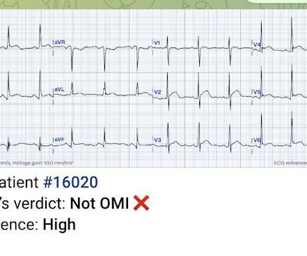
Occlusion myocardialinfarction is a clinical diagnosis Written by Willy Frick (@Willyhfrick). Recall from this post referencing this study that "reciprocal STD in aVL is highly sensitive for inferior OMI (far better than STEMI criteria) and excludes pericarditis, but is not specific for OMI." Circulation , 130 (25). Worrall, C.,
Specific cardiovascular diseases, such as acute myocardialinfarction, arrhythmias, pulmonary hypertension and pericarditis, were also pointed. SiO2 exposure was linked to an increased risk of myocardialinfarction, with potential mechanisms involving inflammation and platelet activation.
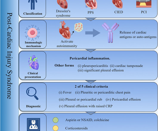
Post-Cardiac Injury Syndrome (PCIS) refers to a collective term encompassing post-myocardialinfarction syndrome, post-pericardiotomy syndrome, and post-traumatic pericarditis. With an aging population, the incidence of PCIS is on the rise annually.
Chronic inflammation plays a role in the pathogenesis of cardiovascular diseases, including atherosclerosis, pericarditis, stroke, and myocardialinfarction (MI).
But there was some doubt as to whether it might be pericarditis because of the ST elevation in I and II, without ST depression in III. Add that to "sharp" pain and a 33 year old, and it is easy to convince yourself that this is, indeed, pericarditis. This is a good sign for myocardialinfarction and does not happen in pericarditis.
This is a value typical for a large subacute MI, n ormal value 48 hours after myocardialinfarction is associated with Post-Infarction Regional Pericarditis ( PIRP ). As already mentioned, this patient could have post-infarction regional pericarditis from a large completed MI. Hammill SC. Edwards WD.
ST depression in lead AVL differentiates inferior ST-elevation myocardialinfarction from pericarditis. Ischemic ST-segment depression maximal in V1-4 (versus V5-6) of any amplitude is specific for occlusion myocardialinfarction (versus nonocclusive ischemia). Am J Emerg Med 2016 5. Meyers et al. Herman et al.
Traditionally used as an anti-inflammatory for pericarditis (inflammation of the lining of the heart), it has recently been shown to result in fewer major heart events in those with a recent heart attack. 7 Secondary prevention following myocardialinfarction: a clinical update. It is an easy win, frequently missed.
When there is MI extending all the way to the epicardium (transmural), that infarcted epicardium is often inflamed (postinfarction regional pericarditis, or PIRP). What complication is the patient with post-infarction regional pericarditis at risk for? One should be on the alert for myocardial rupture. Lessons : 1.
When there is MI extending all the way to the epicardium (transmural), that infarcted epicardium is often inflamed (postinfarction regional pericarditis, or PIRP). In addition, when there is full thickness infarction, especially with inflammation, the myocardium is at risk of "rupture." 3) Oliva et al. (3) Armstrong PW et al.
PR depression, which suggests pericarditis 4. We also showed that, of 47 cases of pericarditis with ST elevation, none had ST depression in aVL. ) The goal of the present analysis was to examine whether the presence of tachycardia identified patients unlikely to have type 1 myocardialinfarction.
Then the patient's pain then resolved spontaneously after 2 sublingual nitroglycerine and another ECG was recorded ECG 2 at 16 minutes ST ELEVATION CONSISTENT WITH INJURY, PERICARDITIS, OR EARLY REPOLARIZATION Overread same Smith : The T-waves are now MUCH smaller. The S-wave is reconstituted. The inferior findings are much less pronounced.
Appearance of abnormal Q waves early in the course of acute myocardialinfarction: implications for efficacy of thrombolytic therapy. MYOCARDIAL RUPTURE AND POSTINFARCTION REGIONAL PERICARDITIS KEY POINTS · Myocardial rupture occurs in 1 to 1.5% Myocardial Rupture and Postinfarction Pericarditis.
Answer: "It does not look like myocardialinfarction". ECG Diagnosis: Normal variant ST Elevation vs. Pericarditis. Uncertain whether there is pericarditis or normal variant. No objective signs of pericarditis (no rub, no effusion, no positional pain) 3. What do you think? He sent the prehospital ECG.
These latter findings are typical of pericarditis, but pericarditis never has reciprocal ST depression. The ECG is diagnostic of occlusion myocardialinfarction (OMI). Usually with pericarditis and myocarditis — hyperacute T waves (HATW) are not present. S mith : there is STE in lead III and reciprocal STD in aVL.
Primary adverse events were defined as myocardialinfarction, thromboembolism, transient ischemic attack, diaphragmatic paralysis, pneumothorax, heart block, pulmonary edema, vagal nerve injury, pericarditis, major vascular access complication or bleeding, death, stroke, or any other cerebrovascular accident.
First, many on Twitter said "Pericarditis". This is NOT pericarditis, which virtually NEVER has ST depression any where except aVR. See our publication: ST depression in lead aVL differentiates inferior ST-elevation myocardialinfarction from pericarditis There is STE in inferior leads, high lateral leads, and V4-V6.
This is a bad ST vector orientation, because it causes widespread STE and one of the most important mistakes that needs to be avoided here is thinking of the diagnosis of pericarditis. Such an out-of-proportion STE is virtually never seen in pericarditis. Considerations on the naming of myocardialinfarctions. 2019.09465.
As myocardialinfarction (MI) and many other diagnoses (for example left ventricular hypertrophy, prior MI etc.) We are happy to announce that our "OMI Toolbox" application has just released and ready for your use. can cause ST-segment elevation (STE) on electrocardiogram (ECG), the distinction between them may be hard and complicated.
I do not think this is acute occlusion myocardialinfarction (OMI). There is also mild pericardial enhancement consistent with pericarditis. QTc's were 330 ms and 373 ms This is what I texted back: These look like they are a very pronounced case of Benign T-wave Inversion. Get an emergent contrast echocardiogram.
So in anterior leads, for diagnosis of ST elevation myocardialinfarction, V1, the cutoff is usually 2 mm, while 1 mm is enought in other leads. PR segment elevation and depression can occur in atrial infarction and pericarditis. When there is ST depression, even 0.5 mm is enough, to consider abormal ST.
You do NOT see this in normal variant STE, nor in pericarditis. Cardiac Troponin Changes to Distinguish Type 1 and Type 2 MyocardialInfarction and 180-Day Mortality Risk. Here is the computer interpretation: (Veritas algorithm) This is what I said: "This is diagnostic of an acute inferior MI. Murakami M.
The initial computer and final cardiology interpretation was a differential: “ST elevation, consider early repolarization, pericarditis, or injury.” Hyperacute T waves can be a useful sign of occlusion myocardialinfarction if appropriately defined. But STEMI criteria ignore all this and look at ST segments in isolation.
Differential of peri-infarct pericardial fluid The differential includes 1) pericarditis with effusion or 2) hemopericardium. Differential of peri-infarct pericardial fluid The differential includes 1) pericarditis with effusion or 2) hemopericardium. Myocardial rupture is not uncommon.
Clinical questions : Is this an occlusion myocardialinfarction and does the patient need the cath lab? Prominent J waves and ventricular fibrillation caused by myocarditis and pericarditis after BNT162b2 mRNA COVID-19 vaccination. The relationship between J wave and ventricular tachycardia during Takotsubo cardiomyopathy.
The exception is with postinfarction pericarditis , in which a completed transmural infarct results in inflammation of the subepicardial myocardium and STE in the distribution of the infarct, and which results in increased STE and large upright T-waves. These findings together are more commonly seen with pericarditis.
We organize all of the trending information in your field so you don't have to. Join thousands of users and stay up to date on the latest articles your peers are reading.
You know about us, now we want to get to know you!
Let's personalize your content
Let's get even more personalized
We recognize your account from another site in our network, please click 'Send Email' below to continue with verifying your account and setting a password.


Let's personalize your content