This site uses cookies to improve your experience. To help us insure we adhere to various privacy regulations, please select your country/region of residence. If you do not select a country, we will assume you are from the United States. Select your Cookie Settings or view our Privacy Policy and Terms of Use.
Cookie Settings
Cookies and similar technologies are used on this website for proper function of the website, for tracking performance analytics and for marketing purposes. We and some of our third-party providers may use cookie data for various purposes. Please review the cookie settings below and choose your preference.
Used for the proper function of the website
Used for monitoring website traffic and interactions
Cookie Settings
Cookies and similar technologies are used on this website for proper function of the website, for tracking performance analytics and for marketing purposes. We and some of our third-party providers may use cookie data for various purposes. Please review the cookie settings below and choose your preference.
Strictly Necessary: Used for the proper function of the website
Performance/Analytics: Used for monitoring website traffic and interactions
This comprehensive evaluation included the use of ultrasound echocardiograms, computed tomography (CT) scans, electrocardiograms, mutagenesis analysis, and structural analysis to gain insights into the patient's condition and the underlying mechanisms of PD.
The patient's laboratory studies revealed troponin mildly elevated at 25 ng/L but liver enzymes, lipase were normal. Gallbladder ultrasound was negative for stones. Here is the EM decision making: "The patient's EKG revealed some repolarization abnormalities but no clear signs of a STEMI. Chest x-ray was normal.
The 3DCT scan was able to predict the calcium arc (P<0.0001) and minimal lumen area by intravascular ultrasound (P=0.003).CONCLUSIONS:Preprocedural The mean amount of procedural contrast (P<0.0001), mean radiation (P=0.03), and median procedure time (P=0.03) were significantly lower in the intervention group.
We report our experience validating this large animal model for translational and preclinical research.Methods3 animal experiments were performed at the UCLA TRIC laboratory between 02/2022 and 10/2022. The animals were transferred to the catheter laboratory and image guidance was performed with a monoplane Siemens Artis Zeego.
These may include the utilization of ultrasound guidance for arterial access, optimization of sheath and catheter selection based on patient anatomy, and the adoption of patent hemostasis devices to minimize complications and enhance overall procedural outcomes.
These may include the utilization of ultrasound guidance for arterial access, optimization of sheath and catheter selection based on patient anatomy, and the adoption of patent hemostasis devices to minimize complications and enhance overall procedural outcomes.
Discussing further, Catheterization Laboratory, also called Cath Lab, is a medical examination room where angiogram, angioplasty, ablation, and implantation of pacemaker are carried out. Ultrasound, TEE, and IVUS play an influential role in dropping the usage of angiographic imaging. Building Smart Cath Labs is highly related to it.
Department of Laboratory Medicine, Hennepin County Medical Center; Professor, University of Minnesota School of Medicine Stephen W. Lee, MD – Department of Emergency Medicine, Hennepin County Medical Center, Minneapolis, MN Yader Sandoval, MD - Department of Cardiovascular Medicine, Mayo Clinic, Rochester, MN. Apple, Ph.D.
Transcript of the video: Echocardiography is now not restricted to the echocardiographic laboratory. During echocardiography, a transducer transmits the ultrasound beam towards the heart. It is used in the emergency department, at bedside, in the intensive care unit as well as in the operating room.
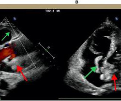
A bedside cardiac ultrasound was performed with a parasternal long axis view demonstrated below: There is a large pericardial effusion with collapse of the right ventricle during systole. Electrical alternans — was first observed in the laboratory by Herring in 1909. This patient is only pseudo-stable. She has already had syncope.
Residents aged 18 years or older in the sampled areas were included in this study, and data were collected through questionnaires, physical examinations, laboratory tests, carotid ultrasound examinations, and biological sample collection. 2021-KY-1289-001).Results:Among respectively.
The final bit of bad news relates to the common laboratory and imaging tests cardiologists typically use to assess for heart dysfunction. Unfortunately, the objective data tracks these symptoms.
Bedside ultrasound showed no effusion and moderately decreased LV function, with B-lines of pulmonary edema. Be certain that your laboratory value is accurate and that it corresponds with the ECG findings! Angio had shown some acute disease in the saphenous vein graft to the posterior descending artery off of the RCA.
Check : [vitals, SOB, Chest Pain, Ultrasound] If the patient has Abdominal Pain, Chest Pain, Dyspnea or Hypoxemia, Headache, Hypotension , then these should be considered the primary chief complaint (not syncope). Aortic Dissection, Valvular (especially Aortic Stenosis), Tamponade. Good History and Physical exam, including a.
The patient was given aspirin 325 mg and laboratory workup was initiated. Nevertheless, the operator performed intravascular ultrasound and saw erupted calcium nodule consistent with plaque erosion. Initial high sensitivity troponin I (hsTnI) was 41 ng/L (reference: 35 ng/L). Echocardiogram showed inferior hypokinesis.
Case continued A bedside cardiac ultrasound revealed grossly preserved left ventricular function, no appreciable wall motion abnormality, pericardial effusion, or obvious valvular abnormality. The terminal part of the T-wave is inverted in lead III, and reciprocally terminally upright in lead aVL.
We organize all of the trending information in your field so you don't have to. Join thousands of users and stay up to date on the latest articles your peers are reading.
You know about us, now we want to get to know you!
Let's personalize your content
Let's get even more personalized
We recognize your account from another site in our network, please click 'Send Email' below to continue with verifying your account and setting a password.


Let's personalize your content