Remove Laboratory Remove Pulmonary Remove Ultrasound
article thumbnail

AngioJet thrombectomy with extracorporeal membrane oxygenation support for an acute large-scale pulmonary embolism with bilateral atrial thrombosis: a case report of catastrophic antiphospholipid syndrome

Frontiers in Cardiovascular Medicine

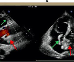
Notably, acute massive pulmonary embolism (PE) with bilateral atrial thrombosis is an exceptional occurrence in CAPS. Acute pulmonary embolism (PE) is a common cardiovascular disease that progresses rapidly and has a high mortality rate. It primarily affects small vessels, seldom impacting large vessels.

article thumbnail

Interplay between mitochondrial dysfunction and lysosomal storage: challenges in genetic metabolic muscle diseases with a focus on infantile onset Pompe disease

Frontiers in Cardiovascular Medicine

This comprehensive evaluation included the use of ultrasound echocardiograms, computed tomography (CT) scans, electrocardiograms, mutagenesis analysis, and structural analysis to gain insights into the patient's condition and the underlying mechanisms of PD. Further genetic testing identified a homozygous mutation c.2662G>T

article thumbnail

Chest Pain and Inferior ST Elevation.

Dr. Smith's ECG Blog

Department of Laboratory Medicine, Hennepin County Medical Center; Professor, University of Minnesota School of Medicine Stephen W. While sensitivity of this sign is very low — its presence is highly suggestive of longstanding and severe pulmonary disease. Apple, Ph.D. Clin Cardiol 22:334-344, 1999 ).

article thumbnail

A woman in her 20s with syncope

Dr. Smith's ECG Blog

A bedside cardiac ultrasound was performed with a parasternal long axis view demonstrated below: There is a large pericardial effusion with collapse of the right ventricle during systole. Electrical alternans — was first observed in the laboratory by Herring in 1909. This patient is only pseudo-stable. She has already had syncope.

article thumbnail

Basics of Echocardiography

All About Cardiovascular System and Disorders

Transcript of the video: Echocardiography is now not restricted to the echocardiographic laboratory. During echocardiography, a transducer transmits the ultrasound beam towards the heart. The aorta, right ventricular outflow tract and pulmonary artery up to its bifurcation is imaged in the upward angulation shown in the left panel.

article thumbnail

Emergency Department Syncope Workup: After H and P, ECG is the Only Test Required for Every Patient.

Dr. Smith's ECG Blog

Check : [vitals, SOB, Chest Pain, Ultrasound] If the patient has Abdominal Pain, Chest Pain, Dyspnea or Hypoxemia, Headache, Hypotension , then these should be considered the primary chief complaint (not syncope). Aortic Dissection, Valvular (especially Aortic Stenosis), Tamponade. Good History and Physical exam, including a.