This site uses cookies to improve your experience. To help us insure we adhere to various privacy regulations, please select your country/region of residence. If you do not select a country, we will assume you are from the United States. Select your Cookie Settings or view our Privacy Policy and Terms of Use.
Cookie Settings
Cookies and similar technologies are used on this website for proper function of the website, for tracking performance analytics and for marketing purposes. We and some of our third-party providers may use cookie data for various purposes. Please review the cookie settings below and choose your preference.
Used for the proper function of the website
Used for monitoring website traffic and interactions
Cookie Settings
Cookies and similar technologies are used on this website for proper function of the website, for tracking performance analytics and for marketing purposes. We and some of our third-party providers may use cookie data for various purposes. Please review the cookie settings below and choose your preference.
Strictly Necessary: Used for the proper function of the website
Performance/Analytics: Used for monitoring website traffic and interactions
The ECG in Figure-1 — was obtained from a middle-aged woman with positional tachycardia and diaphoresis with change of position from suprine to sitting. My THOUGHTS on the ECG in Figure-1: The rhythm is sinus tachycardia at ~105/minute ( ie, The R-R interval is regular — and just under 3 large boxes in duration ).
ischemia) or it can be secondary to abnormal depolarization (e.g Discussion: The ECG in today's case does not have typical ST depression vector of diffuse subendocardial ischemia. The ST vector in subendocardial ischemia (SEI) usually has the largest amount of ST depression in leads II and V5, towards the apex of the heart. (ST
She had a single chamber ICD/Pacemaker implanted several years prior due to ventricular tachycardia. Are you confident there is no ischemia? Answer : The ECG above shows a regular wide complex tachycardia. Said differently, the ECG shows a rather slow ventricular tachycardia with a 2:1 VA conduction. Is this: 1.
A prehospital 12-lead was recorded: There is a regular wide complex tachycardia. The computer diagnosed this as Ventricular Tachycardia. There is a wide complex regular tachycardia at a rate of 226. Toothache, incidental Wide Complex Tachycardia Could it be fascicular VT or Bundle Branch VT ( i.e., idiopathic VT )?
The ECG shows severe ischemia, possibly posterior OMI. But cardiac arrest is a period of near zero flow in the coronary arteries and causes SEVERE ischemia. It takes time for that ischemia to resolve. The patient was brought to the ED and had this ECG recorded: What do you think? And what do you want to do?
Ventricular tachycardia is a potentially life threatening cardiac arrhythmia. On the ECG, ventricular tachycardia can be defined as three or more ventricular ectopic beats occurring in a sequence at a rate more than 100 per minute. Another rare form of ventricular tachycardia is bidirectional ventricular tachycardia.
It should be kept in mind that on occasions, beta-one agonist can result in increased ventricular ectopy e.g., in severe myocardial ischemia (by increasing myocardial demand), or sometimes with congenital long-QT syndrome. Even with tachycardia and a paced QRS duration of ~0.16
Then, a polymorphic ventricular tachycardia occurs over 7 beats. The QT interval of the sinus beats does not appear prolonged, thus ruling out Torsades de Pointes tachycardia. The most likely cause of this type of polymorphic ventricular tachycardia during a stress EKG is cardiac ischemia/coronary artery disease.
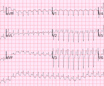
Heart rate/rhythm: consider antidotes for brady/tachy-arrhythmias, and for sinus tachycardia consider fluids for vasodilation and benzodiazepines for agitation. Electrical conduction and axis: consider sodium bicarb for QRS > 100 especially if RBBB or terminal rightward shift, and magnesium for QTc> 500.
At about this point in the process — I like to take a closer LOOK at the 12-lead tracing, to ensure there is no acute ischemia or infarction that might need immediate attention. C ASE C onclusion : I lack detailed follow-up from today's case — other than knowing that the Atrial Tachycardia was controlled.
This is ischemic ST depression, and could be due to increasing tachycardia, with a heart rate over 130, but that is unlikely given that the patient is now complaining of crushing chest pain and that there was tachycardia all along. They agreed ischemia was likely in the setting of demand given DKA and infection.
It is a regular narrow complex tachycardia. There is a lot of ST depression -- this is ischemia caused by the very fast rate and is an indication for emergent electrical cardioversion. Ischemia is gone. Regular Narrow complex tachycardia, if not sinus tach, is AVNRT, AVRT, or atrial flutter with 1:1 or 2:1 conduction.
Three or more ventricular beats in a row at a rate above 100 per minute is termed ventricular tachycardia. Ventricular tachycardia lasting more 30 seconds or requiring termination earlier due to hemodynamic compromise is called sustained ventricular tachycardia. Either case, the treatment is ablation of the right bundle.
Many of the changes seen are reminiscent of LVH with “strain,” and downstream Echo may very well corroborate such a suspicion, but since the ECG isn’t the best tool for definitively establishing the presence of LVH, we must favor a subendocardial ischemia pattern, instead. Type I ischemia. Type II ischemia.
He was rushed by residents into our critical care room with a diagnosis of STEMI, and they handed me this ECG: There is sinus tachycardia with ST elevation in II, III, and aVF, as well as V4-V6. ACS and STEMI generally do not cause tachycardia unless there is cardiogenic shock. He had this ECG recorded. Are the lungs clear?
Here is his initial rhythm strip (it is not a full 10 seconds): Wide complex tachycardia, rate 235 This is a very wide complex regular tachycardia at a rate of 235. It should be considered to be Ventricular Tachycardia and treated as such. He was pale and diaphoretic (in shock) with a thready radial pulse. So what happened?
An Initial ECG was performed: Initial ECG: Sinus tachycardia with prolonged QT interval (QTc of 534 ms by Bazett). She was admitted to the ICU where subsequent ECGs were performed: ECG at 12 hours QTc prolongation, resolution of T wave alternans ECG at 24 hours Sinus tachycardia with normalized QTc interval. No ischemic ST changes.
The above ECGs show the initiation and continuation of a polymorphic ventricular tachycardia. Polymorphic ventricular tachycardia can be ischemic, catecholaminergic or related to QT prolongation. Most such rhythms in the setting of ischemia are VF and will not convert without defibrillation. Acute ischemia?
In any case, the ECG is diagnostic of severe ischemia and probably OMI. So this could be myocarditis but in my opinion needs an angiogram before making that diagnosis. == Dr. Nossen Comment/Interpretation: Evaluation of ischemia on an ECG can be very challenging. Concordant STE of 1 mm in just one lead or 2a.
Here was his initial ED ECG: There is sinus tachycardia at a rate of about 140 There is profound ST Elevation across all precordial leads, as well as I and aVL. I said I think there is a fixed stenosis in the LAD and the tachycardia and stress caused a type 2 STEMI.
While the initial impression might not immediately suggest ventricular tachycardia (VT), a closer examination raises suspicion. Additionally, the qR morphology, particularly in a patient with right bundle branch block (RBBB) type wide QRS complex tachycardia (WQCT), lends further support for VT. What is the rhythm?
DISCUSSION: The 12-lead EKG EMS initially obtained for this patient showed severe ischemia, with profound "infero-lateral" ST depression and reciprocal ST elevation in lead aVR. The ECG cannot diagnose the etiology of ischemia; it only the presence of ischemia, from whatever etiology.
A 50-something male with unspecified history of cardiomyopathy presented in diabetic ketoacidosis (without significant hyperkalemia) with a wide complex tachycardia and hypotension. Analysis: there is a wide complex tachycardia. This was the interpretation I put into the system: WIDE COMPLEX TACHYCARDIA. It is regular.
The ECG shows sinus tachycardia with RBBB and LAFB, without clear additional superimposed signs of ischemia. The Initial ECG in Today's Case: As per Dr. Meyers — the initial ECG in today's case shows sinus tachycardia with bifascicular block ( = RBBB/LAHB ). Sinus Tachycardia ( common in any trauma patient. ).
We have also shown several cases in which atrial flutter hides true, active ischemia. Tachycardia and ST Elevation. Tachycardia to this degree can cause ST segment changes in several ways. Tachycardia to this degree can cause ST segment changes in several ways. Christmas Eve Special Gift!! Is this inferor STEMI?
Here is his ECG: Original image, suboptimal quality Quality improved with PM Cardio digitization The ECG is highly suggestive of acute right heart strain, with sinus tachycardia, S1Q3T3, and T wave inversions in anterior and inferior with morphology consistent with acute right heart strain. Moreover, there is tachycardia.
Generally, the condition is considered benign but can be associated with ischemia. Myocardial bridging (MB) is an inborn abnormality where a segment of an epicardial coronary artery dives transiently into myocardium resulting in systolic arterial compression that normalizes during diastole.
During active chest pain an ECG was recorded: Meyers ECG interpretation: Sinus tachycardia, normal QRS complex, STD in V2-V6, I, II, III and aVF. Learning Points: You must learn and recognize the ECG patterns of OMI and subendocardial ischemia to best understand the ECG in acute care medicine.
Sinus tachycardia has many potential causes. This is especially true for the elderly patient with sinus tachycardia. What is the cause of the sudden tachycardia? She had a very elevated troponin T at 12,335 ng/L at the time of presentation. The patient in today’s case suddenly became tachycardic while sleeping.
Instead, antiarrhythmic drugs such as amiodarone or ß-blockers may be needed — and/or treatment targeted to correcting ischemia. Although IV Mg++ is also indicated as initial treatment of PMVT with a normal QT — it is clearly less likely to respond to IV Mg++, than when the QT interval is prolonged.
Dr Jones is known for his Master Classes in Advanced ECG Interpretation and his published texts, Getting Acquainted With Wide Complex Tachycardias, Getting Acquainted With Laddergrams, and Getting Acquainted With Ischemia and Infarction. His books are available on Amazon.com and on BarnesAndNoble.com.
There is a narrow complex tachycardia at a rate of 130. ECGs: there is a regular narrow complex tachycardia still at a rate of exactly 130, with no P-waves and also no change since the prehospital ECG. During tachycardia, in this case, the baseline continuously undulates especially in leads II, aVR and V1; very good for atrial flutter.
Precordial ST depression may be subendocardial ischemia or posterior STEMI. Notice there is tachycardia. I have warned in the past that one must think of other etiologies of ischemia when there is tachycardia. V4-V6, is much more likely to be posterior than subendocardial ischemia. There is no ST elevation.
This usually represents posterior OMI, but in tachycardia and especially after cardiac arrest, this could simply be demand ischemia, residual subendocardial ischemia due to the low flow state of the cardiac arrest. This rules out subendocardial ischemia and is diagnostic of posterior OMI. V4-5 continue to show STD.
Pediatric exercise testing may be used for evaluation of various disorders of cardiac rhythm rather than for inducible ischemia as in adults. Ventricular arrhythmias during exercise can be documented in congenital long QT syndromes as well as in catecholaminergic polymorphic ventricular tachycardia.
Is there ischemia? ECG Diagnosis is either : 1) junctional rhythm with new LBBB, and possibly ischemia 2) accelerated idoventricular rhythm with possible ischemia, and possibly related to restoration of normal perfusion. Exactly how they relate to ischemia, chest pain, and reperfusion can only be speculated about.
Whenever you see tachycardia with bundle branch block, you should suspect that it is rate related BBB. After resolution, there was T-wave inversion in V1-V3, highly suggestive of ischemia. There are features of the T-wave inversion, however, which argue against ischemia. Moreover, and importantly, there was sinus tach.
This proves effective treatment of the recurrent ischemia. The patient had no further symptoms of ischemia. EKG 3 is diagnostic for developing re-occlusion, and EKG 4 proves that the nitrates relieved the ischemia. = This proves effective treatment of the recurrent ischemia." Here was her final EKG prior to discharge.
It should be known that each category can easily manifest the generic subendocardial ischemia pattern. In general, subendocardial ischemia is a consequence of global supply-demand mismatch that usually ameliorates upon addressing, and mitigating, the underlying cause. What’s interesting is that the ECG can only detect ischemia.
This point is particularly relevant regarding ECG #2 — because sinus tachycardia is seen on this earlier ECG. ECG Blog #184 — illustrates the "magical" mirror-image opposite relationship with acute ischemia between lead III and lead aVL ( featured in Audio Pearl #2 in this blog post ). Cardiol 27:674-677, 2004 ).
His response: “subendocardial ischemia. Smith : It should be noted that, in subendocardial ischemia, in contrast to OMI, absence of wall motion abnormality is common. With the history of Afib, CTA abdomen was ordered to r/o mesenteric ischemia vs ischemic colitis vs small bowel obstruction. Anything more on history?
Tachycardia and ST Elevation. == MY Comment , by K EN G RAUER, MD ( 7/7 /2024 ): == Among the most rewarding type of case for me during my days working in the ED — would be seeing a patient who presented with acute CP ( C hest P ain ) — who I would be able to “cure” simply by recognizing and treating their arrhythmia. Would you give lytics?
With normal EF the tachycardia is not compensatory. The same reciprocal relationship is seen in severe subendocardial ischemia, just with opposite vector direction where V1 can show ST elevation) Below you can find the 3D model of the heart and coronary vessels. And the patient has sinus tachycardia. ( HR about 90-100/min.
ACUTE MI (I allowed Acute MI to be in the report because I knew there would be an elevated troponin from ischemia, which is the definition of acute MI -- but in this case it would most likely be a Type 2 MI from tachycardia) There is also LA-RA lead reversal. Atrial fib may cause Occlusion mimic."
We organize all of the trending information in your field so you don't have to. Join thousands of users and stay up to date on the latest articles your peers are reading.
You know about us, now we want to get to know you!
Let's personalize your content
Let's get even more personalized
We recognize your account from another site in our network, please click 'Send Email' below to continue with verifying your account and setting a password.


Let's personalize your content