This site uses cookies to improve your experience. To help us insure we adhere to various privacy regulations, please select your country/region of residence. If you do not select a country, we will assume you are from the United States. Select your Cookie Settings or view our Privacy Policy and Terms of Use.
Cookie Settings
Cookies and similar technologies are used on this website for proper function of the website, for tracking performance analytics and for marketing purposes. We and some of our third-party providers may use cookie data for various purposes. Please review the cookie settings below and choose your preference.
Used for the proper function of the website
Used for monitoring website traffic and interactions
Cookie Settings
Cookies and similar technologies are used on this website for proper function of the website, for tracking performance analytics and for marketing purposes. We and some of our third-party providers may use cookie data for various purposes. Please review the cookie settings below and choose your preference.
Strictly Necessary: Used for the proper function of the website
Performance/Analytics: Used for monitoring website traffic and interactions
In this phenomenon, a thrombus forms within the lumen of the stent graft component of the frozen elephant trunk prosthesis and puts the patient at risk for downstream embolization with visceral or lower limb ischemia. Therefore, the presence of ILT is associated with increased short-term mortality and morbidity.
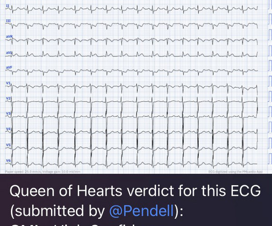
This EKG is diagnostic of transmural ischemia of the inferior wall. If it is angina, lowering the BP with IV Nitroglycerine may completely alleviate the pain and the (unseen) ECG ischemia. Transmural ischemia (as seen with the OMI findings on ECG) is not very common with demand ischemia, but is possible. Smith SW. .
Precordial ST depression may be subendocardial ischemia or posterior STEMI. I have warned in the past that one must think of other etiologies of ischemia when there is tachycardia. The OM-1 was opened and stented, then the LAD was stented 3 days later. There is no ST elevation. How can we tell the difference?
A 39 yo otherwise healthy man with no riskfactors was walking at the mall when he developed chest pressure. Thus, there are some suspicious abnormalities, but no definite signs of ischemia. The difference is significant and highly suggests posterior ischemia. He was diaphoretic. It is very subtle but real.
5 ICSS‐ MRI study (International Carotid Stenting Study Magnetic Resonance Imaging Study), indicated that patients with periprocedural hemodynamic depression had decreased cerebral blood flow and increased the risk of new lesions in imaging.6 Additional riskfactors (delirium vs no delirium) included a history of stroke (17.5%/7.8%),
The patient’s angiogram should have been expedited, but the EKG change was not recognized as recurrence of transmural ischemia. RAO Caudal View Post PCI This is the RAO Caudal view after thrombectomy and stent placement. The conventional computer algorithm called “ sinus tachycardia, otherwise normal EKG ”.
She had zero CAD riskfactors. Now you have ECG and troponin evidence of ischemia, AND ventricular dysrhythmia, which means this is NOT a stable ACS. The lesion was stented. It they are static, then they are not due to ischemia. This is better evidence for ischemia than any other data point.
He had multiple cardiovascular riskfactors and the EM physician strongly suspected ACS. They found 100% acute mid-LAD Occlusion MI, stented with excellent angiographic result. Written by Pendell Meyers A male in his early 50s presented with waxing and waning chest pain starting at rest. mm of the "required" 1.0
In other words, the inferior ST segments in the first ECG show more straightening which is more concerning for ischemia. The culprit lesion was opened and stented. That said — in this older man with riskfactors and new CP, ST-T wave changes in leads aVL and in the inferior leads suffice to suggest acute OMI until proven otherwise.
RiskFactors: High Cholesterol. Angiogram soon after (around 4 hrs after presentation) showed multi vessel CAD, with culprit lesion total occlusion of the first obtuse marginal branch (OM1), which was stented. So, I'm a follower of your blog, and I think I have a interesting case that I attended yesterday." Vitals Signs: Normal."
He has no cardiovascular riskfactors except smoking for 10 pack-years. Compare to the anatomy after stenting: The lower of the 2 now easily seen branches is the circumflex, now with excellent flow. Case A 39-year-old male without prior medical history presents with chest pain that started 2 hours prior to presentation.
PCI is commonly used to open blocked arteries to treat significant myocardial ischemia , which occurs when the heart muscle does not get enough oxygenated blood. During PCI, an operator inserts a stent into a blocked artery through a catheter in the groin or arm.
One of the most common questions I get is, “ Do I need a stent to treat my heart disease?” ” Typically, several of this person’s friends have had stents, so it seems natural to ask. First, we must understand what a stent is and why it is used. The stent ‘unblocks’ it. Flow is restored.
He had no apparent riskfactors for cardiovascular disease. This proves that the first one was, surprisingly, due to ischemia!! He was successfully treated with one drug eluting stent. At the time of arrival to the ED, the patient reported 1/10 chest pain with normal vital signs. A prior ECG was available for comparison.
I still use CT CAC scans for risk estimation, but I am always conscious of their limitations, and I never use it in the setting of suspected obstructive coronary artery disease. Share But why do a CT if riskfactor modification is the priority? 5 ISCHEMIA Research Group. Eur Heart J. 2020 Jan 14;41(3):407-477.
This was stented. If there is polymorphic VT with a long QT on the baseline ECG, then generally we call that Torsades, but Non-Torsades Polymorphic VT can result from ischemia alone. I have read articles that say that patients without ischemia are at low risk of complications from hypokalemia, But it is not entirely without risk.
Women also had more cardiovascular riskfactors, including hypertension (66.6% The diagnosis typically requires classic clinical features, with no evidence of obstructive coronary disease, and typical findings of ischemia on functional studies. versus 63.2%; P <0.001), hyperlipidemia (68.9% years of age versus 59.0±8.4
In this case, the context is a 51 year old man with riskfactors presenting with acute onset substernal chest pain with nausea and vomiting. The operator performed intravascular ultrasound and visualized acute plaque rupture with thrombus formation and placed a stent. The pre-EKG probability for OMI could hardly be much higher!
Like, viability, scars, futility, and benefits of revascularization, imaging-assisted PCI, impact of PCI on exercise capacity, importance of riskfactor management, etc. otherwise, if you keep getting even the slightest doubt and anxiety over the hidden blocks, go for a stent immediately at a good Institution. (My
Case submitted by Andrew Grimes, Advanced Care paramedic, with additions from Jesse McLaren and Smith An 84-year-old male with a notable cardiac history (CABG, multiple stents) woke at 0500hrs with pressure in his chest, diaphoresis, and light-headedness. He had a 100% RCA occlusion which was stented.
We organize all of the trending information in your field so you don't have to. Join thousands of users and stay up to date on the latest articles your peers are reading.
You know about us, now we want to get to know you!
Let's personalize your content
Let's get even more personalized
We recognize your account from another site in our network, please click 'Send Email' below to continue with verifying your account and setting a password.


Let's personalize your content