Remove Ischemia Remove Risk Factors Remove STEMI
article thumbnail

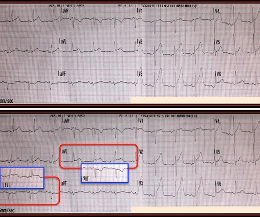
An undergraduate who is an EKG tech sees something. The computer calls it completely normal. How about the physicians?

Dr. Smith's ECG Blog

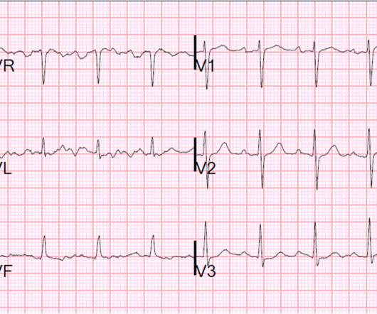
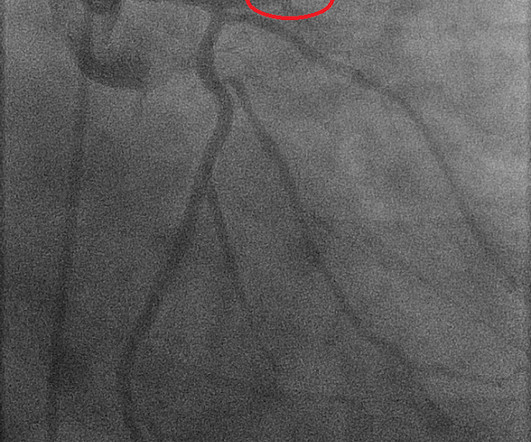
This EKG is diagnostic of transmural ischemia of the inferior wall. If it is angina, lowering the BP with IV Nitroglycerine may completely alleviate the pain and the (unseen) ECG ischemia. Transmural ischemia (as seen with the OMI findings on ECG) is not very common with demand ischemia, but is possible. Smith SW. .

article thumbnail

A Non STEMI that needs the cath lab now.

Dr. Smith's ECG Blog

He had some cardiac risk factors including hypertension, on meds, but no previous coronary disease. He had an immediate ED ECG: There is artifact, but the findings appear to be largely gone now The diagnosis is acute MI, but not STEMI. There is about 1 mm of STE in aVR I con sidered but rejected subendocardial ischemia.

STEMI 52
article thumbnail

Is this acute STEMI? LV Aneurysm? Would you give Thrombolytics?

Dr. Smith's ECG Blog

No risk factors, leads a healthy lifestyle. See my formula for differentiating anterior LV aneurysm (that is to say, persistent ST elevation after old MI) from acute anterior STEMI. Both support acute anterior STEMI. No risk factors, leads healthy lifestyle. BP 112/80, SpO2 100%. PCI is not an option."

article thumbnail

5 Cardiologists said this is not a STEMI. But was it an OMI?

Dr. Smith's ECG Blog

He had multiple cardiovascular risk factors and the EM physician strongly suspected ACS. Over the next few hours, four other general cardiologists "signed off on the initial ECG without recognizing STEMI." Learning Points: STEMI criteria misses 25-40% of OMI, like this case for example. mm of the "required" 1.0

STEMI 52
article thumbnail

Precordial ST depression. What is the diagnosis?

Dr. Smith's ECG Blog

Precordial ST depression may be subendocardial ischemia or posterior STEMI. If you thought it might be a posterior STEMI, then you might have ordered a posterior ECG [change leads V4-V6 around to the back (V7-V9)]. I have warned in the past that one must think of other etiologies of ischemia when there is tachycardia.

STEMI 52
article thumbnail

What does the angiogram show? The Echo? The CT coronary angiogram? How do you explain this?

Dr. Smith's ECG Blog

This suggests further severe ischemia. See "Prevention of cardiovascular disease events in those with established disease (secondary prevention) or at very high risk".) This has resulted in an under-representation of STEMI MINOCA patients in the literature. Detailed coronary artery evaluation not performed. Downstream vasospasm?

article thumbnail

Missed Acute MI, with coronary occlusion, evidence only by T-wave inversion in V2 and evolving ST depression in V3

Dr. Smith's ECG Blog

A 39 yo otherwise healthy man with no risk factors was walking at the mall when he developed chest pressure. Thus, there are some suspicious abnormalities, but no definite signs of ischemia. The difference is significant and highly suggests posterior ischemia. He was diaphoretic. It is very subtle but real.