This site uses cookies to improve your experience. To help us insure we adhere to various privacy regulations, please select your country/region of residence. If you do not select a country, we will assume you are from the United States. Select your Cookie Settings or view our Privacy Policy and Terms of Use.
Cookie Settings
Cookies and similar technologies are used on this website for proper function of the website, for tracking performance analytics and for marketing purposes. We and some of our third-party providers may use cookie data for various purposes. Please review the cookie settings below and choose your preference.
Used for the proper function of the website
Used for monitoring website traffic and interactions
Cookie Settings
Cookies and similar technologies are used on this website for proper function of the website, for tracking performance analytics and for marketing purposes. We and some of our third-party providers may use cookie data for various purposes. Please review the cookie settings below and choose your preference.
Strictly Necessary: Used for the proper function of the website
Performance/Analytics: Used for monitoring website traffic and interactions
Theres ST elevation in V3-4 which meets STEMI criteria, which could be present in either early repolarization, pericarditis or injury. Lets see what happens in the current STEMI paradigm. Emergency physician: STEMI neg but with elevated troponin = Non-STEMI The first ECG was signed off. What do you think?
Cardiogenic shock (CS)is the most feared event following STEMI. We tend to perceive CS as an exclusive complication of STEMI. The incidence is half of that of STEMI, i.e., 2.5-5%. might show little elevation with considerable overlap of left main STEMI vs NSTEMI ) 2.Onset ACS pathophysiology is not that simple.
This suggests further severe ischemia. Contemporary research studies of MINOCA have evaluated the prognosis of these patients, reporting a 12-month all-cause mortality of 4.7% (95% confidence interval, 2.6–6.9), This has resulted in an under-representation of STEMI MINOCA patients in the literature. Downstream vasospasm?
STE limited to aVR is due to diffuse subendocardial ischemia, but what of STE in both aVR and V1? The last section is a detailed discussion of the research on aVR in both STEMI and NonSTEMI. Cardiac arrest can cause diffuse subendocardial ischemia, usually transient (it often resolves as time goes by after ROSC).
Here is his ECG: There is no clear evidence of OMI or ischemia. Moreover, the research which appears to confirm this idea was indeed in relation to the circumflex, but they did not study Occlusion ; rather, they studied asymptomatic coronary disease. A 40-something male with no previous cardiac disease presented with chest pain.
The receiving emergency physician consulted with interventional cardiology who stated there was no STEMI. Is there STEMI? Possible mechanisms of ventricular arrhythmias elicited by ischemia followed by reperfusion. Circulation Research , 56 (2), 184–194. The patient continued having chest pain. What is the rhythm?
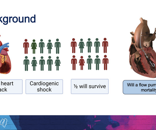
The new trial, called DanGer Shock , is the first trial powered to examine whether the use of micro-axial flow pumps can improve survival in ST-elevation myocardial infarctions (STEMI, the most serious type of heart attack) that are complicated by cardiogenic shock. Overall, we have more complications, but we also save lives.”
Smith15, and PERFECT Study Author Group 1 Hennepin County Medical Center, 2 Minneapolis Medical Research Foundation, 3 Background : The Smith-modified Sgarbossa criteria (MSC) are frequently recommended for diagnosing acute coronary occlusion (ACO; STEMI-equivalent) in the setting of ventricular paced rhythm (VPR). Singer14, Stephen W.
It is equivalent to a transient STEMI. Now you have ECG and troponin evidence of ischemia, AND ventricular dysrhythmia, which means this is NOT a stable ACS. It they are static, then they are not due to ischemia. This is better evidence for ischemia than any other data point. Again, cath lab was not activated.
Contemporary research studies of MINOCA have evaluated the prognosis of these patients, reporting a 12-month all-cause mortality of 4.7% (95% confidence interval, 2.6–6.9), This has resulted in an under-representation of STEMI MINOCA patients in the literature. From Gue at al.
I remember Allie well from her days in the Research volunteer program at Hennepin. 2) The STE in V1 and V2 has an R'-wave and downsloping ST segments, very atypical for STEMI. Cardiology was consulted and they agreed that the EKG had an atypical morphology for STEMI and did not activate the cath lab. Bicarb 20, Lactate 4.2,
I do research on Cardiologs' algorithm: Smith SW et al. But lead V2 has a worrisome amount of ST elevation, and in a chest pain patient, I would be worried about STEMI. A Deep Neural Network learning algorithm outperforms a conventional algorithm for emergency department electrocardiogram interpretation.
2 days later This is a typical LVH pattern, without ischemia Patient underwent 4 vessel CABG. In the setting of prior stenting and reduced left ventricular ejection fraction, would pursue a heart team revascularization approach Syntax score 28.5,
There’s minimal concave ST elevation in III which does not meet STEMI criteria, so this ECG is "STEMI negative". Use STEMI criteria to identify acute coronary occlusion: the ECG was STEMI negative 2. A repeat ECG was done on way to cath lab: "STEMI negative" again. The cath lab was activated. Take home 1.
He is a STEMI patient (1 year old) with mild LV dysfunction and thinning of IVS and anterior wall. Finally, we can grow a potential research hypothesis. Every PCI, by default, is perceived as good by our flawed coronary intellect. A single patient experience Let me share a patient consult from a remote town of north India.
It is a long read, meant only for those who want to know the hidden intricacies in the concept of “Time window” in STEMI and its important Implication in patient care. [08/11, 08/11, 12:13] Dr S Venkatesan: Time windows are related to time taken for myocardial cell death because of ischemia. How can they be different? [08/11,
It is possible there is microvascular dysfunction producing residual transmural ischemia. But this is most common when there is prolonged ischemia, and this patient had the fastest reperfusion imaginable! Circulation Research , 114 (12), 18521866. Here is the final angiogram following placement of a stent in the ostial RCA.
We organize all of the trending information in your field so you don't have to. Join thousands of users and stay up to date on the latest articles your peers are reading.
You know about us, now we want to get to know you!
Let's personalize your content
Let's get even more personalized
We recognize your account from another site in our network, please click 'Send Email' below to continue with verifying your account and setting a password.


Let's personalize your content