This site uses cookies to improve your experience. To help us insure we adhere to various privacy regulations, please select your country/region of residence. If you do not select a country, we will assume you are from the United States. Select your Cookie Settings or view our Privacy Policy and Terms of Use.
Cookie Settings
Cookies and similar technologies are used on this website for proper function of the website, for tracking performance analytics and for marketing purposes. We and some of our third-party providers may use cookie data for various purposes. Please review the cookie settings below and choose your preference.
Used for the proper function of the website
Used for monitoring website traffic and interactions
Cookie Settings
Cookies and similar technologies are used on this website for proper function of the website, for tracking performance analytics and for marketing purposes. We and some of our third-party providers may use cookie data for various purposes. Please review the cookie settings below and choose your preference.
Strictly Necessary: Used for the proper function of the website
Performance/Analytics: Used for monitoring website traffic and interactions
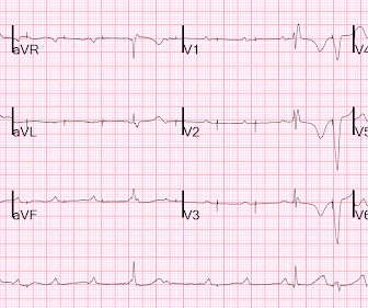
She also has sick sinus syndrome (SSS) and intermittent high grade AV block for which she had a dual chamber pacemaker implanted. ECG#1 Assessing ischemia on an ECG with wide QRS complexes (AIVR, ventricular pacing, BBB, etc) can be challenging. In the ECG above there are several features indicative of ongoing transmural ischemia.
She had a single chamber ICD/Pacemaker implanted several years prior due to ventricular tachycardia. Are you confident there is no ischemia? Primary VT , and the VT with tachycardia is causing ischemia with chest discomfort (supply-demand mismatch/type 2 MI)? The last echocardiography 12 months ago showed HFmrEF.
Place temporary pacemaker 3. It should be kept in mind that on occasions, beta-one agonist can result in increased ventricular ectopy e.g., in severe myocardial ischemia (by increasing myocardial demand), or sometimes with congenital long-QT syndrome. See this post: How a pause can cause cardiac arrest 2.
I tell the residents: "The pacemaker is just common sense: if there is no beat, it provides one; if there is one, it keeps itself from pacing." This is similar to Ken Grauer's comment at the bottom: "What would I do if I were a pacemaker?" This made me realize that pacemaker function is not as well understood as I thought.
Is a pacemaker needed? As a result — IF no "fixable" cause is found ( ie, ischemia/infarction — electrolyte disturbance — rate-slowing medication ) — then because of the AV block and very slow heart rate, this patient will probably need a pacemaker. QUESTIONS: HOW would you interpret the rhythm in Figure-1 ?
The fact that R waves 2 through 6 are junctional does make ischemia more difficult to interpret -- but not impossible. Back to the assessment of ischemia: Returning to the ECG, the leads that catch my eye first are -- I, II, V4, V5, V6. Ischemia can be disguised by a wide escape rhythm, which decreases the sensitivity of ECG.
T wave alternans is characterized by variation in T-wave morphology in the setting of consistent pacemaker and QRS morphology. (1) Alternation in ST segment appearance ( or in the amount of ST elevation or depression ) — is often linked to ischemia. Teaching Points: 1.
There is no definite evidence of acute ischemia. (ie, Simply stated — t he patient was having recurrent PMVT without Q Tc prolongation, and without evidence of ongoing transmural ischemia. ( Some residual ischemia in the infarct border might still be present. Both episodes are initiated by an "R-on-T" phenomenon.
We admitted him for probable EP study and possible pacemaker. He underwent pacemaker placement and is doing fine. SSS is by far the most common reason for permanent pacemaker placement. These include rate-slowing medication recent ischemia/infarction hypothyroidism sleep apnea. Learning Points: 1.
There was no evidence of ischemia. She had a permanent pacemaker implanted. After pacer AND conversion to sinus rhythm: Computer diagnosis: IMPRESSION ELECTRONIC VENTRICULAR PACEMAKER ABNORMAL RHYTHM ECG What is missing from this interpretation? We are not told how ischemia has been ruled out in this case. Hyperkalemia.
My Immediate Impression — was that this elderly woman with a several week history of symptoms would most likely leave the hospital with a pacemaker. This suggests ischemia of uncertain duration. A permanent pacemaker was placed. PEARL # 2: Interpretation of the 12-lead ECG in Figure-1 is no easy task!
Again, see Ken's discussion below) Discussion continued The absence of pace spikes suggests this is not a pacemaker/ICD-related rhythm in this patient with an ICD. Are the apparent P-waves (which now we suspect might not be P-waves) actually part of the QRS, in which case the QRS is even wider than it appears? Where does the QRS begin?
Edits by Meyers and Smith A man in his 70s with PMH of hypertension, hyperlipidemia, type 2 diabetes, CVA, dual-chamber Medtronic pacemaker, presented to the ED for evaluation of acute chest pain. EKG shown here: LAFB with no clear signs of OMI or ischemia. Sent by Pete McKenna M.D. Triage ECG: What do you think?
Automatic activity refers to enhanced pacemaking function (typically from a non sinus node source), for example atrial tachycardia. Possible mechanisms of ventricular arrhythmias elicited by ischemia followed by reperfusion. The most common triggered arrhythmia is Torsades de Pointes. And is there new left bundle branch block (LBBB)?
Isoprenalin was discontinued, and a temporary transveous pacemaker was implanted. The patient stabilized following pacemaker placement. Extensive conduction system abnormalities can have various causes (ischemia, genetic, infectious, amyloid, etc).
Poor blood supply Ischemia, or inadequate blood supply to the heart, is an abnormality that can be detected in an ECG test. ECG testing is also carried out to see how medicines work during treatment and the pacemaker's functioning. If the vital organs do not get their blood supply back quickly, it can lead to death.
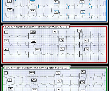
My thoughts were the following: ECGs #1 and #2 showed a completely unreliable sinus pacemaker, with presumed high-grade 2nd-degree AV block — and frequent resultant pauses of over 2 seconds ( that would have been even longer had it not been for intermittent relief from the atrial escape focus ). What Does this All Mean?
100% occluded RCA with TIMI 0 flow Post drug-eluting stent placement with TIMI 3 flow While in the cath lab, she transiently developed complete heart block and became hypotensive requiring transvenous pacemaker placement and transient pressors. The transvenous pacemaker was removed the following day and pressors were not required again.
Some patients with pacemakers and metal prosthesis can’t be exposed to such strong magnetic fields. These are therefore not looking for coronary disease but instead ischemia heart disease. Remember functional tests tell you about ischemia and anatomical tests tell you about coronary disease.
The medics recorded the following initial ECG at time 0: The computer read (see below) gives no further comment beyond ventricular pacemaker. There is no way the ST-T wave should change from lead V4-to-V5 as we see here given the similar all negative QRS appearance in these 2 leads, unless there is acute ischemia. What do you think?
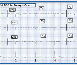
Learning Points: Ectopic atrial rhythm can produce atrial repolarization findings that can be confused for acute ischemia, STEMI, or OMI. If you can safely and easily increase the patient's heart rate, you can convert the patient to sinus and repeat the ECG to see if the atrial repolarization wave was the cause of the concern for ischemia.
These findings are concerning for inferior wall ischemia with possible posterior wall involvement. It is also not a wandering pacemaker — because change in atrial pacing site is gradual with that disorder. The morphology in V2 is also concerning and it appears that the ST segment is being pushed down, as in ST depression.
I initially suspected V2 as being placed too high on the chest, but there is no accompanying inverted P wave here, so the positioning is sound. It’s important to stress the presence of a normal QRS (i.e.,
Therefore, she underwent temporary pacemaker placement and overdrive pacing at a rate of 90 bpm to keep the heart rate up in order to prevent these PVCs triggering ventricular arrhythmia. Instead, antiarrhythmic drugs such as amiodarone or ß-blockers may be needed — and/or treatment targeted to correcting ischemia. Acute ischemia?
Rapid Fire Challenging Structural Heart Imaging Cases with Heart Team Panel; Follow-up of Pacemakers and ICDS for the Non-electrophysiologist; The Real Reasons Your Patient with Heart Failure was Readmitted: Noncardiac Comorbidities, Geriatric Syndromes and Social Determinants of Health; and Death by a Thousand Cuts!
Such findings would normally suggest primary ischemia with concomitant surveillance of coronary occlusion, but these ST/T changes might very well be secondary to the Escape mechanism at hand. He received a permanent pacemaker during the subsequent inpatient stay. Hospital transport was unremarkable.
Ruling out other potential causes of bradycardia ( ie, recent ischemia-infarction; hypothyroidism ). I would bet that this patient will soon receive a permanent pacemaker. = Follow-Up: Our interpretation of this Holter was passed on to the patient's primary physician.
These include: i ) Use of rate-slowing medication ( ie, ß-blockers, digoxin, verapamil/diltiazem, etc. ) ; ii ) Acute or recent infarction or ischemia; iii ) Hypothyroidism; iv ) Neurologic injury; v ) Electrolyte disturbance; and , vi ) Sleep apnea. I therefore thought the significance of this finding in today’s case was uncertain.
No evidence of ischemia. Many favored pacemaker implantation at this time. Ultimately ( with completely informed patient consent ) — the decision was made to implant a pacemaker. CASE Disposition: Opinions of consulting cardiologists on this case were divided.
Evidence of acute ischemia (may be subtle) vii. Negative predictors of adverse outcome: Pacemaker Pre-syncope or "near-syncope," but there is still some small risk (5, 18) These last two are identified in studies, but I consider them dangerous signs and symptoms in their own right, as above: 10. Left BBB vi. Pathologic Q-waves viii.
No evidence for ischemia jumps out. How does a pacemaker accomplish RBBB morphology? Quick aside on device terminology (feel free to skip): A "single chamber" pacemaker is a device with only one lead. A "dual chamber" pacemaker is a device with an atrial lead and a ventricular lead. ECG 1 What do you think?
Unless a potentially fixable cause of this rhythm can be found ( ie, acute ischemia from recent infarction; electrolyte disturbance, use of rate-slowing medication ) the patient will need a pacemaker. This strongly suggests ischemia which could be recent and a contributing ( or precipitating) cause of todays abnormal rhythm.
PEARL # 2: Despite the challenge of assessing a ventricular rhythm for underlying ischemia and/or infarction there are primary ST-T wave changes that are seen in Figure-3 that suggest an ongoing acute event. The above said Today's patient had other ideas. He remained remarkably stable despite the above conduction disorder.
It is possible there is microvascular dysfunction producing residual transmural ischemia. But this is most common when there is prolonged ischemia, and this patient had the fastest reperfusion imaginable! Here is the final angiogram following placement of a stent in the ostial RCA. In the midst of this, she went into VF.
We organize all of the trending information in your field so you don't have to. Join thousands of users and stay up to date on the latest articles your peers are reading.
You know about us, now we want to get to know you!
Let's personalize your content
Let's get even more personalized
We recognize your account from another site in our network, please click 'Send Email' below to continue with verifying your account and setting a password.


Let's personalize your content