This site uses cookies to improve your experience. To help us insure we adhere to various privacy regulations, please select your country/region of residence. If you do not select a country, we will assume you are from the United States. Select your Cookie Settings or view our Privacy Policy and Terms of Use.
Cookie Settings
Cookies and similar technologies are used on this website for proper function of the website, for tracking performance analytics and for marketing purposes. We and some of our third-party providers may use cookie data for various purposes. Please review the cookie settings below and choose your preference.
Used for the proper function of the website
Used for monitoring website traffic and interactions
Cookie Settings
Cookies and similar technologies are used on this website for proper function of the website, for tracking performance analytics and for marketing purposes. We and some of our third-party providers may use cookie data for various purposes. Please review the cookie settings below and choose your preference.
Strictly Necessary: Used for the proper function of the website
Performance/Analytics: Used for monitoring website traffic and interactions
ObjectiveThe prevalence of acute myocardialinfarction, a severe ischemic cardiac disease, is on the rise annually. Over the past two decades, studies related to angiogenesis in acute myocardialinfarction have increased rapidly. The collection of literature was gathered using the Web of Science Core Collection database.
These methods only alleviate symptoms of heart failure and myocardialischemia and improve patients' quality of life by partially restoring myocardial reperfusion. The main content of this paper is to explore the application of stem cells and gene technology in the treatment of myocardialinfarction (MI).
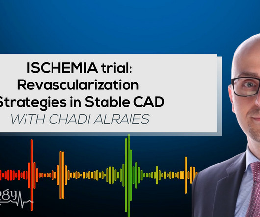
In the ISCHEMIA (International Study of Comparative Health Effectiveness with Medical and Invasive Approaches) trial, researchers examined the risk of ischemic events in patients with stable coronary artery disease. years, with 57.1% occurring within 30 days after CABG. Original article: Redfors B et al.
This confirms that the pain was ischemia and is now resovled. Thus, it has recently become generally accepted that most plaque ruptures resulting in myocardialinfarction occur in plaques that narrow the lumen diameter by 40% of the arterial cross section may be involved by plaque. The i nitial hs troponin I returned 75%.
Myocardialinfarction (MI) is a deadly disease. It can cause serious myocardial ischemic necrosis due to coronary occlusion. However, reperfusion itself could induce more severe injury, called myocardialischemia/reperfusion injury (MI/RI).
The largest trial to examine the impact of colchicine in acute myocardialinfarction (MI) found that both acute and long-term colchicine use did not reduce cardiovascular death, myocardialinfarction, stroke, or ischemia-driven revascularization.
The goal of the MINT trial was to compare the effect of a liberal vs. restrictive blood transfusion strategy on acute myocardialinfarction (MI) in patients with concomitant anemia.
No ischemia. This is a conundrum, because it is clear that the patient is having an acute MI, the ECG is dynamic, but the pain is very mild and there is no ECG evidence of active transmural ischemia. We already know that the ischemia is ongoing, though mild (because of the persistent pain). Q-waves are even more pronounced.
BackgroundStrain assessed by cardiac magnetic resonance (CMR) is a key prognostic indicator in myocardialinfarction. However, the strain characteristics and prognostic value in myocardialinfarction with nonobstructive coronary arteries (MINOCA) with different causes are unclear.
In the heart, autophagy is regulated mainly through mitophagy due to the metabolic changes of cardiomyocytes caused by ischemia and hypoxia. Myocardial remodeling is characterized by gradual heart enlargement, cardiac dysfunction, and extraordinary molecular changes.
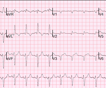
The ECG did not meet STEMI criteria, and the final cardiology interpretation was “ST and T wave abnormality, consider anterior ischemia”. Hence the first ECG was labeled 'anterior ischemia' based on ST depression, rather than identifying this as reciprocal from posterior OMI. But are there any other signs of Occlusion MI? Meyers et al.
Such T-waves are almost always reciprocal to ischemia in the region of aVL (although aVL looks n ormal here) , and in a patient with chest pain are nearly diagnostic of ischemia. Ischemia on the ECG can be very subtle and is easily missed. Did the ECG offer unseen hints? Lesson : 1.
Both acute and long-term colchicine use to treat patients with acute myocardialinfarction (MI) did not reduce cardiovascular death, myocardialinfarction, stroke or ischemia-driven revascularization.
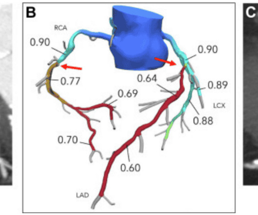
6 This novel study marks a significant milestone in the field, evaluating the effectiveness of FFR CT in detecting ischemia-producing coronary stenosis in patients with severe PAD. Diagnosis and treatment of ischemia-producing coronary stenoses improves 5-year survival of patients undergoing major vascular surgery.” 101272, [link].
BackgroundSlow flow/no-reflow (SF-NR) during percutaneous coronary intervention (PCI) is associated with poor prognosis of patients with acute myocardialinfarction (AMI). Currently, effective treatment is not available for SF-NR. However, its effects on SF-NR in the AMI patients during PCI are not clear.
There is akinesis of the distal septum, anterior, apex, and distal inferior wall consistent with LAD territory ischemia or infarction. How large is the infarct? It is impossible to conclude from this that the infarct was VERY large, though it likely was very large since the time to intervention was long.
The 2 coprimary outcomes were target lesion revascularization and myocardialinfarction. The secondary outcomes included ischemia-driven target lesion revascularization, target vessel myocardialinfarction, death, cardiac death, target vessel revascularization, stent thrombosis, and major adverse cardiac events.
If this STD were due to LVH or to subendocardial ischemia, rather than posterior OMI, it would be maximal in V5 and V6. If I saw this without the STD V2-V4, I would not make anything of it, and even with that precordial STD, I am not convinced that it is a manifestation of ischemia. This is a HUGE myocardialinfarction.
Important point: when there is diffuse subendocardial ischemia but no OMI, a wall motion abnormality will not necessarily be present. They agreed ischemia was likely in the setting of demand given DKA and infection. That this is all demand ischemia is unlikely. Lung exam showed diffuse B lines bilaterally. Aslanger's pattern.
There may be ischemia present, but it is not evident on the ECG. In this paper, Dr. Birnbaum writes: "In patients with ACS without LVH, ST depression with negative T waves in the lateral leads is a sign of sub-endocardial ischemia and is an independent predictor of adverse outcome [11 – 13]. Notice the S-wave in V2 is 45 mm.
Advances in cardiovascular imaging have improved the ability to identify coronary artery stenosis in patients with KD, yet knowledge gaps remain regarding optimal frequency of serial imaging and the best imaging modality to identify those at risk for inducible myocardialischemia.
This EKG is diagnostic of transmural ischemia of the inferior wall. Smith: note also the terminal QRS distortion in lead III (absence of S-wave without a prominent J-wave). . __ Smith comment 1 : the appropriate management at this point is to lower the blood pressure (lower afterload, which increases myocardial oxygen demand).
Myocardialischemia may induce myocardial fibrosis, a condition that progressively leads to ventricular remodeling, heightening the risk of heart failure. The timely detection of myocardial fibrosis is crucial for intervention and improved outcomes. The results demonstrated tracer-specific uptake (SUVmax = 4.6)
In some cases the ischemia can be seen "through" the flutter waves, whereas in other cases the arrhythmia must be terminated before the ischemia can be clearly distinguished. First , there can simply be diffuse ST depressions (which obligates reciprocal STE in aVR) associated with tachycardia which are not indicative of ischemia.
Abstract: N(6)-methyladenosine (m6A) methylation modification is involved in the progression of myocardialinfarction (MI). The ischemia/reperfusion (I/R) injury mouse model and hypoxia/reoxygenation (H/R) cell model were established. The results provided a theoretical basis that ALKBH5 is a potential target for MI treatment.
Martha Gulati, MD, director of preventive cardiology in the department of cardiology at Los Angeles-based Cedars-Sinai's Smidt Heart Institute has raised awareness of two heart conditions needing better diagnostic tools ischemia with no obstructive coronary arteries and myocardialinfarction with no obstructive coronary arteries.
Background Untreated multivessel disease (MVD) in acute myocardialinfarction (AMI) has been linked to a higher risk of recurrent ischemia and death within one year. The immediate non-IRA PCI is associated with a significantly lower occurrence of unplanned ischemia-driven PCI (OR 0.60; confidence interval [CI] 0.44–0.83)
V5-V6) of any amplitude, is specific for Occlusion MyocardialInfarction (vs. non-occlusive ischemia) Ongoing ischemic symptoms in NSTEMI is already an indication for emergent cath, regardless of the ECG. Sometimes posterior leads help, and sometimes they falsely reassure.
This AI technology can detect 35 cardiac determinations (14 arrhythmias and 21 morphologies), including serious conditions like acute myocardialinfarction ( MI ) and the most common types of cardiac ischemia, using a reduced leadset. KAI 12L employs multiple deep neural network algorithms, trained and validated on more than 1.75
FFRCT analysis revealed high rates of asymptomatic lesion-specific coronary ischemia (65%), severe ischemia (52%), multivessel ischemia (36%), and left main ischemia (8%). Meanwhile, the usual care group’s coronary ischemia statuses remained unknown (because they didn’t test for it).
Myocardialinfarction is among the top causes of mortality worldwide. Infarct-related torsade de pointes (TdP) is an uncommon complication. In the context of myocardialinfarction, coronary artery bypass graft (CABG) surgery is the prevalent therapeutic modality associated with several early and late complications.
A 53-year-old male presenting emergently with signs of myocardialinfarction received immediate coronary angiography and thoracic CT-scan showing occlusion of the first marginal coronary branch without possibility of revascularization and minimal pericardial extravasation.
See these 2 articles Association between pre-hospital chest pain severity and myocardial injury in ST elevation myocardialinfarction: A post-hoc analysis of the AVOID study Author links open overlay panel [link] 1 Background We sought to determine if an association exists between prehospital chest pain severity and markers of myocardial injury.
IntroductionAcute spinal cord ischemia syndrome (ASCIS) is a rare disease that is thought to comprise roughly only 1.2% The mechanism is thought to be multifactorial due vasospasm, cerebral vasculitis, vascular thrombosis, cardioembolism from cocaine‐induced myocardialinfarction or cardiomyopathy, and hypertensive surges [9].
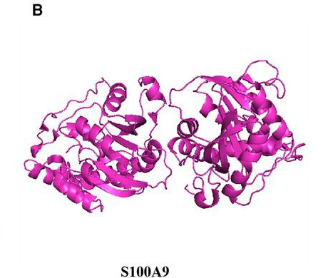
This article evaluates the utility of S100A8/A9 protein as a biomarker and therapeutic target for diagnosing cardiovascular diseases, considering its structural features, fundamental biological properties, and its multifaceted influence on cardiovascular conditions including atherosclerosis, myocardialinfarction, myocardialischemia/reperfusion injury, (..)
BackgroundFerroptosis, an iron‐dependent form of regulated cell death, is a major cell death mode in myocardialischemia reperfusion (I/R) injury, along with mitochondrial permeability transition‐driven necrosis, which is inhibited by cyclosporine A (CsA). Journal of the American Heart Association, Ahead of Print.
The first ECG was labeled “anterior subendocardial ischemia”, but subendocardial ischemia does not localize. If there were diffuse ischemic STD, with precordial STDmaxV5-6 and reciprocal STE-aVR, this would be non-specific subendocardial ischemia from ACS or supply-demand mismatch.
This is a value typical for a large subacute MI, n ormal value 48 hours after myocardialinfarction is associated with Post-Infarction Regional Pericarditis ( PIRP ). Mechanical complications secondary to myocardialinfarction are infrequent due to most patients receiving revascularization quite rapidly.
This suggests diffuse subendocardial ischemia. However, along with that subendocardial ischemia, there is also STE in lead III with reciprocal ST depression in aVL, and some STE in V1. If there is also subendocardial ischemia, the ST depression vector remains leftward, with a reciprocal ST Elevation vector also to the right.
For those who depend on echocardiogram to confirm the ECG findings of ischemia, this should be sobering. In this case, the duration of ischemia was so brief that there was no such evolution, and there was near-normalization. Wellens' syndrome represents a state of reperfusion of the infarct related artery 2. Lessons: 1.
ET Main Tent (Hall B1) This session offers more insights from key clinical trials presented at ACC.24 24 and find out what it all means for your patients.
Cardiovascular disease is the most common cause of death and disability globally, largely driven by myocardialinfarction and ischemic stroke caused by atherosclerosis (plaque build-up in the arteries).
The primary endpoint was the one-year rate of target vessel failure, the composite occurrence of either cardiac death, target-vessel myocardialinfarction, or ischemia-driven target-vessel revascularization.
We organize all of the trending information in your field so you don't have to. Join thousands of users and stay up to date on the latest articles your peers are reading.
You know about us, now we want to get to know you!
Let's personalize your content
Let's get even more personalized
We recognize your account from another site in our network, please click 'Send Email' below to continue with verifying your account and setting a password.


Let's personalize your content