
Unlocking Precision in Interventional Cardiology: The Vital Role of OCT and IVUS
ADN Center of Excellence
NOVEMBER 20, 2023
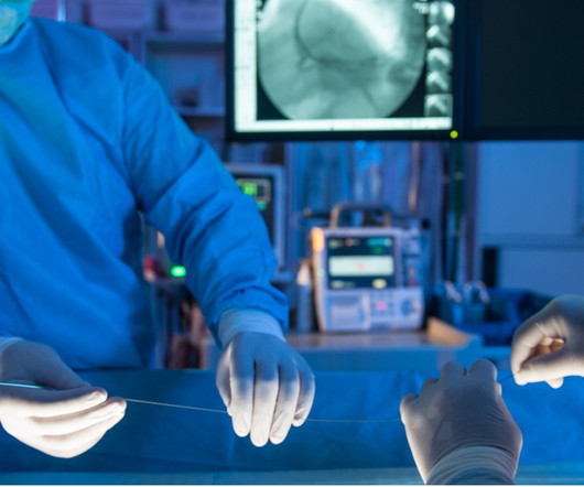
In the realm of interventional cardiology, the quest for precision is an ongoing pursuit. OCT employs light waves to capture high-resolution images, providing intricate details of vessel structures and plaque composition. Advancements in OCT and IVUS constantly refine the approach to diagnosis, treatment, and patient care.


Let's personalize your content