This site uses cookies to improve your experience. To help us insure we adhere to various privacy regulations, please select your country/region of residence. If you do not select a country, we will assume you are from the United States. Select your Cookie Settings or view our Privacy Policy and Terms of Use.
Cookie Settings
Cookies and similar technologies are used on this website for proper function of the website, for tracking performance analytics and for marketing purposes. We and some of our third-party providers may use cookie data for various purposes. Please review the cookie settings below and choose your preference.
Used for the proper function of the website
Used for monitoring website traffic and interactions
Cookie Settings
Cookies and similar technologies are used on this website for proper function of the website, for tracking performance analytics and for marketing purposes. We and some of our third-party providers may use cookie data for various purposes. Please review the cookie settings below and choose your preference.
Strictly Necessary: Used for the proper function of the website
Performance/Analytics: Used for monitoring website traffic and interactions
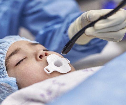
Cardiovascular ultrasound has played a key role in the evolution of early diagnosis of structural heart disease, led by a technology pioneered by Philips: the ‘transesophageal echocardiography’ (TEE) ultrasound transducer. TEE helps cardiologists by providing highly detailed images of the heart and its internal structures.
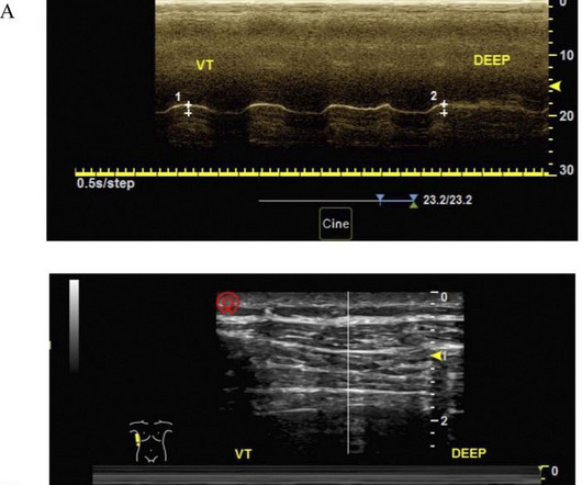
ObjectiveApproximately 10%–70% of patients may develop diaphragmatic dysfunction after cardiac surgery, which may lead to delayed weaning from mechanical ventilation, increased ICU stays, postoperative hospitalization stays, and respiratory complications. However, its impact on prognosis and risk factors remain controversy.
Bedside cardiac ultrasound showed moderately decreased LV function. CASE CONTINUED She was admitted to the ICU. (And of course Ken's comments at the bottom) An elderly obese woman with cardiomyopathy, Left bundle branch block, and chronic hypercapnea presented hypoxic with altered mental status. She was intubated.
Our AI-enabled portfolio, including our Command Center Software Platform, Edison True PACS , and Venue Family ultrasound systems with Caption Guidance , is designed to directly address these issues. "At GE HealthCare, we understand the critical challenges healthcare providers face, from staffing shortages to complex workflows.
He was requiring supplemental oxygen and an initial bedside cardiac ultrasound was unremarkable. He was administered a therapeutic dose of low-molecular weight heparin and transferred to the ICU. Extensive clot burden in bilateral lower extremities was visualized on ultrasound. Cardiology was consulted.
Cannulation is performed in a mobile ICU under mechanical CPR, with ultrasound-guided cannulation as the first choice. Cannulation was performed percutaneously under ultrasound guidance, with an average cannulation time of 9.5 Additionally, witness presence, bystander CPR, and blood gas analysis are considered.
The patient was upgraded to the ICU for closer monitoring. Cardiac Ultrasound may be a surprisingly easy way to help make the diagnosis Answer: pulmonary embolism. Now another, with ultrasound. Echocardiogram showed severe RV dilation with McConnell’s sign and an elevated RVSP. What is the Diagnosis? This is a quiz.
Once stabilized, intravascular ultrasound showed significant thrombus and plaque in the LAD. While preparing for transport to the cardiac ICU, the Impella device malfunctioned, and function could not be restored. This was treated with a drug-eluting stent, but TIMI 3 flow was not achieved.
Course : A CT of the head, neck, chest, abdomen and pelvis showed no other unanticipated injuries and she was admitted to the ICU. Hemodynamic instability in trauma is usually due to bleeding, but if ultrasound shows poor contractility, then this may be due to cardiac contusion.
Given her risk factors (HTN, HLD, ESRD from diabetes) I decided to obtain a broad cardiac workup for the patient: serial ECGs, labs, serial troponins, CXR and bedside cardiac ultrasound. Ultrasounds can be very helpful in guiding your diagnostic pathway: location of WMA on US led to obtaining posterior leads.
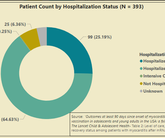
In summary, we have a CDC follow-up study that shows 25% of survey responders with vaccine myocarditis were admitted to the ICU, and one of these cases required a modified type of heart/lung bypass machine to stay alive. But a good long-term prognosis related to these cardiac scars is what everyone hopes for, not what anyone knows.
Smith comment: Point of Care ultrasound is not adequate to rule out wall motion abnormality; moreover, diffuse subendocardial ischemia often has no wall motion abnormality because the epicardium is still contracting. Academic Emergency Medicine 27(S1): S220. Abstract 556.
So I immediately left the room to get an ultrasound machine. While calling for some help and arranging to have her transported to our critical care zone, I got this quick ultrasound which confirmed my suspicion: This quick view was all I was able to obtain in the circumstances.
His ED cardiac ultrasound (which is not at all ideal for detecting wall motion abnormalities, and is also very operator dependent for this finding) was significant for depressed global EF. Fortunately, he was extubated several days later in the ICU with intact baseline mental status and was discharged shortly thereafter to subacute rehab.
in the ICU but survived with excellent function. Beware a negative Bedside ultrasound. The team was notified and they ordered a stat aortagram which showed type A aortic dissection from the aortic valve to the iliacs. Not surprisingly the cardiology HPI changed yet again in the next note following diagnosis of the aortic dissection: ".chest
She was resuscitated and admitted to ICU for presumed sepsis. If you do not have an arterial line, use bedside ultrasound to verify myocardial contractility corresponds to pacing. If you don't have ultrasound (but you should), then palpate a pulse! Her family had not heard from her and called EMS.
We organize all of the trending information in your field so you don't have to. Join thousands of users and stay up to date on the latest articles your peers are reading.
You know about us, now we want to get to know you!
Let's personalize your content
Let's get even more personalized
We recognize your account from another site in our network, please click 'Send Email' below to continue with verifying your account and setting a password.


Let's personalize your content