This site uses cookies to improve your experience. To help us insure we adhere to various privacy regulations, please select your country/region of residence. If you do not select a country, we will assume you are from the United States. Select your Cookie Settings or view our Privacy Policy and Terms of Use.
Cookie Settings
Cookies and similar technologies are used on this website for proper function of the website, for tracking performance analytics and for marketing purposes. We and some of our third-party providers may use cookie data for various purposes. Please review the cookie settings below and choose your preference.
Used for the proper function of the website
Used for monitoring website traffic and interactions
Cookie Settings
Cookies and similar technologies are used on this website for proper function of the website, for tracking performance analytics and for marketing purposes. We and some of our third-party providers may use cookie data for various purposes. Please review the cookie settings below and choose your preference.
Strictly Necessary: Used for the proper function of the website
Performance/Analytics: Used for monitoring website traffic and interactions
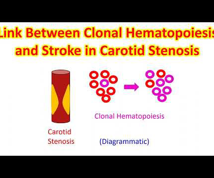
About a fifth of all ischemic strokes are attributed to embolization of ruptured atherosclerotic plaque from carotid arterial stenosis. But it has been difficult to predict which person with asymptomatic carotid artery stenosis is likely to progress to symptomatic carotid disease and stroke. J Am Coll Cardiol. doi: 10.1016/j.jacc.2024.03.389.
This comprehensive literature review focuses on acute stroke related to intracranial atherosclerotic stenosis (ICAS), with an emphasis on ICAS-large vessel occlusion. Various risk factors, including hypertension, diabetes, hyperlipidemia, smoking, and advanced age lead to ICAS, which in turn results in stroke through different mechanisms.
Intubated and given nitric oxide for pulmonary hypertension. Four- week echo continues to show pulmonic valve stenosis. The ECG: In spite of the pulmonary valve stenosis, this ECG is within normal limits for this 4-week old. Weaned in NICU over 10 days. Echocardiogram during that time showed stiff pulmonic valve.
Yasser Sammour, MD, MScpresented "Long-Term Hemodynamic Performance and Clinical Outcomes in Small and Large Aortic Annuli Patients with Severe Aortic Stenosis."This Safety outcomes, including the presence of renal artery stenosis greater than 70%, were also evaluated. "The This study is a pooled analysis from the U.S.
Stroke, Volume 56, Issue Suppl_1 , Page ADP36-ADP36, February 1, 2025. All patients had headache, and funduscopic examination demonstrated papilledema for all patients. All patients had headache, and funduscopic examination demonstrated papilledema for all patients.
Introduction:Medical treatment of internal carotid artery stenosis consists of treatment of underlying conditions such as hypertension, dyslipidemia, and diabetes mellitus, as well as antiplatelet therapy. This may lead the way to new drug therapies for carotid artery stenosis.
Hypertension, Ahead of Print. By now chronic renovascular disease (RVD) due to renal artery stenosis is recognized as a major source of renovascular hypertension and renal disease. Almost a hundred years have passed since obstruction of the renal artery has been recognized to raise blood pressure.
A 63 year old man with a history of hypertension, hyperlipidemia, prediabetes, and a family history of CAD developed chest pain, shortness of breath, and diaphoresis after consuming a large meal at noon. He called EMS, who arrived on scene about two hours after the onset of pain to find him hypertensive at 220 systolic.
Objective A novel artificial intelligence-based phenotyping approach to stratify patients with severe aortic stenosis (AS) prior to transcatheter aortic valve replacement (TAVR) has been proposed, based on echocardiographic and haemodynamic data.
BackgroundRING finger protein 213 (RNF213) p.R4810K is an established risk factor for moyamoya disease and intracranial artery stenosis in East Asian people. Recent evidence suggests its potential association with extracranial cardiovascular diseases, including pulmonary hypertension.
BACKGROUNDHypertension is often codiagnosed in patients with moyamoya disease (MMD), a progressive intracranial steno‐occlusive vasculopathy; this has principally been attributed to renal artery stenosis (up to 10%). Blood pressure measurements and antihypertensive agent use were recorded pre‐ and postoperatively. respectively.
However, the long-term outcomes in patient with an intermediate stenosis received FFR have not yet been investigated comprehensively.Methods:We retrospective included 558 patients underwent both coronary artery angiography (CAG) and FFR. The nomogram consists of age, smoking, hypertension, diabetes mellitus (DM), hyperuricemia, and FFR≤0.8
Bar plots: in red, patients with low flow-low gradient (LF-LG) aortic stenosis; in blue, patients with normal flow-high gradient (HG) aortic stenosis; in black: controls. Aim Cardiac remodelling plays a major role in the prognosis of patients with aortic stenosis (AS) and could impact the benefits of aortic valve replacement.
IntroductionIdiopathic intracranial hypertension (IIH) is a pathology involving an increase in intracranial pressure leading to symptoms including papilledema, tinnitus, and elevated cerebrospinal fluid opening pressure. Ophthalmology consult revealed bilateral papilledema, upon which an MRI and MRV revealed venous stenosis.
Clinical introduction A woman in her 30s, a case of rheumatic mitral stenosis status post balloon mitral valvuloplasty 15 years prior, presented to urgent care with palpitations and dyspnoea for 1 week. Echocardiography demonstrated severe calcific mitral stenosis with pulmonary hypertension.
Background:Intracranial artery stenosis (ICAS) is a progressive pathological process. In multivariable analysis, only hypertension independently predicted stroke occurrence in the asymptomatic ICAS group (adjusted HR 4.06, 95%CI 1.60-10.33, Stroke, Volume 56, Issue Suppl_1 , Page ATP274-ATP274, February 1, 2025. 10.33, P = 0.003).Conclusions:The
Written by Pendell Meyers A man in his late 30s with history of hypertension, tobacco use, and obesity presented to the Emergency Department for acute chest pain which started approximately 3 hours prior to arrival, in the setting of a very stressful situation. Vitals were within normal limits except some hypertension. Which is true.
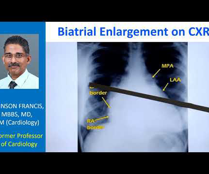
However, underlying lesions such as hypertension, mitral valve disease, COPD, ASD, and TR greatly influence the degree of atrial enlargement. However, in cases of lone AF, AF in hypertension, or chronic AF, both atria tend to dilate equally. In all probability, this dilation is a form of atrial tachycardia and atrial cardiomyopathy.
A 50-something male with hypertension and 20- to 40-year smoking history presented with 1 week of stuttering chest pain that is worse with exertion, which takes many minutes to resolve after resting and never occurs at rest. It is a ssociated with mild dyspnea on exertion. At times the pain does go to his left neck.
Background:Vertebrobasilar artery stenosis (VBAS) can cause posterior circulation strokes (PCS). Hypertension (HTN, 85.4%) and diabetes (DM, 18.9%) were prevalent. Circulation, Volume 150, Issue Suppl_1 , Page A4135852-A4135852, November 12, 2024. Mean age was 69.45 years, with 64.1% Demographics: 69.8% Hispanic, and 5.3% other races.
In this study, we evaluated the relationship between flossing and ICAS, defined as 50% stenosis. The association remained significant after adjustment for age, race, gender, hypertension, diabetes, smoking status, education level and regular dental care use (Adjusted OR 0.61 The log WMH volume was higher (9.50.85
“We are witnessing a paradigm shift in how valvular heart diseases are diagnosed and treated,” said Partho Sengupta , Henry Rutgers Professor of Cardiology and chief of the Division of Cardiovascular Disease and Hypertension at Rutgers Robert Wood Johnson Medical School.
One is ventricular septal defect, second is overriding aorta, third is pulmonary stenosis, usually right ventricular outflow tract stenosis and associated right ventricular hypertrophy. Pulmonary stenosis, which is usually right ventricular outflow tract stenosis. As the name implies, there are four defects.
CardioSignal has already developed digital biomarkers for AFib and heart failure, while more solutions could be on the way for aortic stenosis, coronary artery disease, and pulmonary artery hypertension.
Doppler ultrasonography performed a day after the operation showed an increase in systolic blood velocity, with no observed urine output and raising a suspicion of arterial anastomotic stenosis. The transplant renal artery lesion was intervened with a stent.
I've previously discussed the interesting correlation of a qR pattern in lead V1 in patients with RVH — as strongly suggesting associated pulmonary hypertension ( See ECG Blog #234 and Blog #248 ). The QRS is not wide enough for a complete RBBB — and, lateral limb leads I and aVL both lack terminal S waves.
It was found that all cases of dAVF‐CI exhibited venous hypertension. Additionally, there was a significant association between sinus stenosis and dAVF‐CI (OR: 2.85, 95% CI: 1.16‐7.55, ConclusiondAVFs‐CI tend to occur in relatively young patients and are characterized by the presence of venous hypertension. 7.55, p = 0.027).
FAI was still associated with global CFR after adjusting for traditional risk factors (age, hypertension, diabetes, hyperlipidemia, and smoking). Altogether, FAI can help reveal microcirculatory damage in patients who do not exhibit epicardial artery stenosis.
Results:Among the 36,403 participants (14,676 males, 40.3%), the prevalence of stroke, heart disease, hypertension, diabetes mellitus, and dyslipidemia was 7.4%, 6.4%, 55.7%, 17.3%, and 40.2% 2021-KY-1289-001).Results:Among respectively.
Background:Distinguishing hypertrophic cardiomyopathy (HCM) from other cardiomyopathies with left ventricular hypertrophy (LVH), such as hypertensive LVH, transthyretin amyloid cardiomyopathy (ATTR-CM), and aortic stenosis (AS), is sometimes challenging. Of those, 5 proteins were selected as candidate proteins.
However, CTA head and neck 4 days later demonstrated 90 percent stenosis of the mid left V2 at the C3‐4 level and a 75‐90 percent stenosis of the left mid V2 segment at the C5‐6 level (hard and soft plaque in these areas). He also had moderate stenosis of the right V4 segment.
Yasser Sammour, MD, MScpresented "Long-Term Hemodynamic Performance and Clinical Outcomes in Small and Large Aortic Annuli Patients with Severe Aortic Stenosis."This Safety outcomes, including the presence of renal artery stenosis greater than 70%, were also evaluated. "The This study is a pooled analysis from the U.S.
MRA head demonstrated multifocal arterial stenosis. She was treated with intravenous hydration, permissive hypertension with head of bed in flat position and transferred for further evaluation. MRI brain showed subacute infarcts in left greater than right frontal lobes, corpus collosum and right anterior perforated substance.
So that is why we see straightening of left border, typically heard of in mitral stenosis with left atrial enlargement and mild pulmonary hypertension. When there is gross pulmonary hypertension, instead of these being straight over here, it will form a bulge over here.
Pulmonary hypertension (PH) is a complex and progressive disorder characterised by elevated pulmonary artery pressure. Transcatheter aortic valve implantation (TAVI) is a minimally invasive surgical procedure that has revolutionised the treatment of severe aortic stenosis (AS).
The sample included patients who presented with stroke due to severe stenosis and underwent elective stenting, and those who presented with large vessel occlusion (LVO) with underlying ICAD who underwent rescue stenting following thrombectomy. There were significantly higher incidences in uncontrolled hypertension (28.2%
It was edited by Smith CASE : A 52-year-old male with a past medical history of hypertension and COPD summoned EMS with complaints of chest pain, weakness and nausea. A transthoracic echocardiogram showed an LV EF of less than 15%, critically severe aortic stenosis , severe LVH , and a small LV cavity.
Conclusion:Prior use of RASI for the treatment of hypertension is associated with persistent hypotension after CAS. BP was recorded at frequent intervals for at least 24 hours post-CAS. Blood catecholamine concentrations were assessed in the morning of, and day immediately after, CAS in a subset of patients.
Category 2 : An increase in myocardial oxygen demand due to tachycardia, elevated ventricular afterload (BP or aortic stenosis), or increased wall stretch (admittedly this latter is more complicated) or a decrease in oxygen supply due to hypotension, anemia, hypoxia, or a combination of all of the above. Aortic Stenosis f.
Outflow obstruction leading to venous hypertension was observed in all dAVFs-CI. Sinus stenosis was significantly associated with dAVFs-CI (OR 2.85, 95% CI: 1.16-7.55, Discussion:Venous hypertension is a key angiographic feature dAVFs-CI. 7.55, p = 0.027). 2.05, p < 0.001) and draining veins (OR 2.05, 95% CI 1.05-4.46,
We organize all of the trending information in your field so you don't have to. Join thousands of users and stay up to date on the latest articles your peers are reading.
You know about us, now we want to get to know you!
Let's personalize your content
Let's get even more personalized
We recognize your account from another site in our network, please click 'Send Email' below to continue with verifying your account and setting a password.


Let's personalize your content