This site uses cookies to improve your experience. To help us insure we adhere to various privacy regulations, please select your country/region of residence. If you do not select a country, we will assume you are from the United States. Select your Cookie Settings or view our Privacy Policy and Terms of Use.
Cookie Settings
Cookies and similar technologies are used on this website for proper function of the website, for tracking performance analytics and for marketing purposes. We and some of our third-party providers may use cookie data for various purposes. Please review the cookie settings below and choose your preference.
Used for the proper function of the website
Used for monitoring website traffic and interactions
Cookie Settings
Cookies and similar technologies are used on this website for proper function of the website, for tracking performance analytics and for marketing purposes. We and some of our third-party providers may use cookie data for various purposes. Please review the cookie settings below and choose your preference.
Strictly Necessary: Used for the proper function of the website
Performance/Analytics: Used for monitoring website traffic and interactions
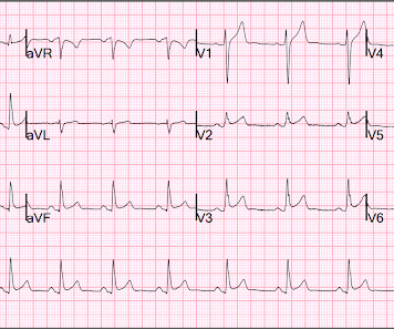
Hospital evaluation for this patient was negative for an acute coronary syndrome ( ie, CT coronary angiogram was normal — troponin was not elevated — and Echo was negative, with no sign of pericardial effusion ). The patient was discharged with a diagnosis of acute pericarditis — and treated with a full course of colchicine and ibuprofen.
This is a 45 yo male who had an inferior STEMI 6 months prior, was found to have severe LAD and left main disease, and was supposed to be set up for CABG a few weeks later, but did not follow up. But it could be anterior STEMI. 40% of anterior STEMI has upward concavity in all of leads V2-V6. is likely anterior STEMI).
She presented to an outside hospital after several days of malaise and feeling unwell. This is a value typical for a large subacute MI, n ormal value 48 hours after myocardial infarction is associated with Post-Infarction Regional Pericarditis ( PIRP ). At the time of admission, her vital signs were normal. Heart rate was in the 80s.
If you were thinking that this is not anterior OMI because there is no reciprocal ST depression , it is important to remember that half of anterior STEMI do NOT have any reciprocal ST depression. Pericarditis? If you were thinking that this is pericarditis, that would be possible in the absence of any clinical information.
These latter findings are typical of pericarditis, but pericarditis never has reciprocal ST depression. Despite active CP — cath lab activation was deferred and this patient was transported to a local hospital without PCI capability. Usually with pericarditis and myocarditis — hyperacute T waves (HATW) are not present.
They informed me that she had just been hospitalized 10 days ago for "some fluid around the heart" and was discharged after one day without incident. Ultimately, she spent several days in the hospital and no further fluid collected. She was diagnosed with pericarditis and spent one day in the hospital without events.
A previously healthy 53 yo woman was transferred to a receiving hospital in cardiogenic shock. So Shark Fin really is just a dramatic representation of STEMI, and can be in any coronary distribution. So this is STEMI, right? Well, don't we see diffuse ST Elevation in Myo-pericarditis (with STD in aVR)? and K was normal.
As always, takotsubo cardiomyopathy and focal pericarditis can mimic OMI, but takotsubo almost never mimics posterior MI, and both are diagnoses of exclusion after a negative cath. The provider contacted cardiology to discuss the case, but cardiology "didn't think it was a STEMI, didn't think he needed emergent cath." Canto et al.
cm diameter in the apex The presence of thrombus led the clinicians to state that this was a "late presentation STEMI." It does take some time for thrombus to form, but the EKG and the troponin profile show that this was NOT a late presentation STEMI. of AMI patients and is often preceded by postinfarction regional pericarditis (PIRP).
for those of you who do not do Emergency Medicine, ECGs are handed to us without any clinical context) The ECG was read simply as "No STEMI." Given his exertional chest pain and elevated troponin, the patient was admitted to the hospital for "NSTEMI" with a plan for left heart catheterization the next day. What is the Diagnosis?
Furthermore, some ECGs may not meet the STEMI criteria but may still be diagnostic for acute coronary occlusion (ACO). Another handy section is for troponin levels and their delta cut-offs, which can be modified according to the kit used in your hospital. We would like to thank Muzaffer Değertekin, MD, PhD, Prof.
This morphology can be cause by or associated with cocaine: A Patient with Cocaine Chest Pain and Prehospital Computer interpretation of STEMI This is OMI of the anterior, lateral, and inferior walls until proven otherwise. But it does not meet STEMI criteria and it was not initially recognized. The cath lab was now activated.
This is a bad ST vector orientation, because it causes widespread STE and one of the most important mistakes that needs to be avoided here is thinking of the diagnosis of pericarditis. Such an out-of-proportion STE is virtually never seen in pericarditis. He arrived to our hospital one hour later.
50% of LAD STEMIs do not have reciprocal findings in inferior leads, and many LAD OMIs instead have STE and/or HATWs in inferior leads instead. The ECG easily meets STEMI criteria in all leads V2-V6, as well. He was then transferred to quaternary care childrens hospital. 24 yo woman with chest pain: Is this STEMI?
He spent almost 2 months in the hospital, and reportedly made a full neurologic recovery. Dyspnea, Chest pain, Tachypneic, Ill appearing: Bedside Cardiac Echo gives the Diagnosis 31 Year Old Male with RUQ Pain and a History of Pericarditis. This patient arrested shortly after hospital arrival. He was prescribed apixaban.
You do NOT see this in normal variant STE, nor in pericarditis. After admission to the hospital, the patient was discharged from the hospital without any investigation of his acute MI. Here is the computer interpretation: (Veritas algorithm) This is what I said: "This is diagnostic of an acute inferior MI.
We organize all of the trending information in your field so you don't have to. Join thousands of users and stay up to date on the latest articles your peers are reading.
You know about us, now we want to get to know you!
Let's personalize your content
Let's get even more personalized
We recognize your account from another site in our network, please click 'Send Email' below to continue with verifying your account and setting a password.


Let's personalize your content