This site uses cookies to improve your experience. To help us insure we adhere to various privacy regulations, please select your country/region of residence. If you do not select a country, we will assume you are from the United States. Select your Cookie Settings or view our Privacy Policy and Terms of Use.
Cookie Settings
Cookies and similar technologies are used on this website for proper function of the website, for tracking performance analytics and for marketing purposes. We and some of our third-party providers may use cookie data for various purposes. Please review the cookie settings below and choose your preference.
Used for the proper function of the website
Used for monitoring website traffic and interactions
Cookie Settings
Cookies and similar technologies are used on this website for proper function of the website, for tracking performance analytics and for marketing purposes. We and some of our third-party providers may use cookie data for various purposes. Please review the cookie settings below and choose your preference.
Strictly Necessary: Used for the proper function of the website
Performance/Analytics: Used for monitoring website traffic and interactions
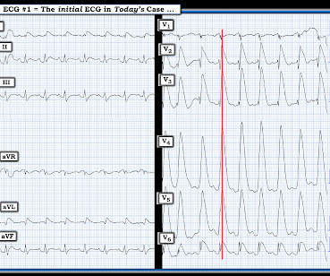
That said — the ECG in Figure-1 should prompt the following considerations: The symmetric chest lead T wave inversion in ECG #1 could be a sign of coronary disease, potentially with acute ischemia. During my decades of working with residents when hospital Attending — by far, the most commonly overlooked vital sign was respiratory rate.
BACKGROUND:Lower-limb amputation rates in patients with chronic limb-threatening ischemia vary across the United States, with marked disparities in amputation rates by gender, race, and income status. 0.98];P=0.019), and those who received care at a safety-net hospital (odds ratio, 0.87 [95% CI, 0.78–0.97];P=0.012) Mean age, 76.6
At the hospital, left main coronary-artery stenosis was seen on angiography (shown in a video). In a 57-year-old man with chest pain, an ECG obtained by EMS showed widespread ST-segment depressions.
The ECG shows severe ischemia, possibly posterior OMI. But cardiac arrest is a period of near zero flow in the coronary arteries and causes SEVERE ischemia. It takes time for that ischemia to resolve. The patient was brought to the ED and had this ECG recorded: What do you think? And what do you want to do?
This EKG is diagnostic of transmural ischemia of the inferior wall. If it is angina, lowering the BP with IV Nitroglycerine may completely alleviate the pain and the (unseen) ECG ischemia. Transmural ischemia (as seen with the OMI findings on ECG) is not very common with demand ischemia, but is possible. Smith SW.
BackgroundLittle is known about treatment variability across US hospitals for patients with chronic limb‐threatening ischemia (CLTI).Methods All patients aged ≥18 years, admitted to nonfederal US hospitals with a primary diagnosis of CLTI, were identified. Journal of the American Heart Association, Ahead of Print.
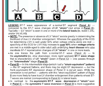
His confusion progressively dissipated enroute to the hospital. Many of the changes seen are reminiscent of LVH with “strain,” and downstream Echo may very well corroborate such a suspicion, but since the ECG isn’t the best tool for definitively establishing the presence of LVH, we must favor a subendocardial ischemia pattern, instead.
ii to show blood flow through the heart muscle and evaluate the presence, extent and degree of myocardial ischemia or infarction. Around 6 million MPI procedures are undertaken each year in theU.S. Flyrcado is now available in selectU.S.markets.
He was treated for infection and DKA and admission to hospital was planned. Important point: when there is diffuse subendocardial ischemia but no OMI, a wall motion abnormality will not necessarily be present. They agreed ischemia was likely in the setting of demand given DKA and infection. 40 mg of furosemide was given.
About 20 minutes later ( on the way to the hospital ) — the patient's CP resolved, and ECG #1 was recorded. ECG Blog #184 — illustrates the "magical" mirror-image opposite relationship with acute ischemia between lead III and lead aVL ( featured in Audio Pearl #2 in this blog post ). ECG Blog #337 — an OMI misdiagnosed as an NSTEMI.
Brittany Weber, MD, PhD , of Brigham and Women’s Hospital, is the 2024 YIA winner for her abstract, "The Frequency, Prevalence, And Outcomes Of Incidentally Detected Coronary Artery Calcium Using Artificial Intelligence Analysis Among Patients With Immune Mediated Inflammatory Diseases.”
DISCUSSION: The 12-lead EKG EMS initially obtained for this patient showed severe ischemia, with profound "infero-lateral" ST depression and reciprocal ST elevation in lead aVR. Author continued : STE in aVR is often due to left main coronary artery obstruction (OR 4.72), and is associated with in-hospital cardiovascular mortality (OR 5.58).
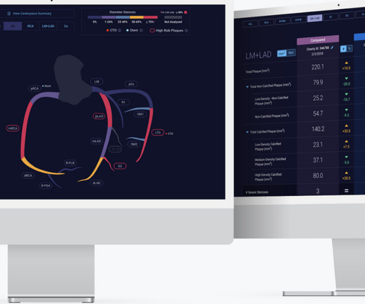
He is a Professor of Radiology and prior to joining Cleerly full-time, served as Vice-Chairman of Radiology at George Washington University (GWU) Hospital in Washington, DC. High Diagnostic Accuracy Of AI-Ischemia in Comparison To PET, FFR-CT, SPECT, and Invasive FFR: A PACIFIC Sub-Study. In Press European Heart Journal CV Imag 2024.This
The patient survived the hospitalization. non-occlusive ischemia) Ongoing ischemic symptoms in NSTEMI is already an indication for emergent cath, regardless of the ECG. A woman in her 50s with chest pain and lightheadedness and "anterior subendocardial ischemia" A man in his 50s with acute chest pain who is lucky to still be alive.
The study was aimed to test the effects of GSNO in ischemia/reperfusion (I/R) Injury through relieving thrombo-inflammation.Methods:Male C57BL/6 mice (n=160) were 10-week-old (20-25 g) at the time of surgery. All animals received humane care in compliance with Beijing Tiantan Hospitals guidance.
My written interpretation on a tracing such as this one would read, "Marked LVH and 'strain' and/or ischemia — with need for clinical correlation." BOTTOM LINE: ECG changes of LV "strain" and/or ischemia that we see on today's initial ECG — were not present 9 years earlier. Please see ECG Blog #73 for additional details ).
The "good news" — is that after an extended hospitalization, the patient was finally discharged home, and doing well. = Shark Fin" ST segment elevation is most often a sign of severe transmural ischemia that results from acute coronary occlusion. The ECG in Figure-1 — was obtained following successful resuscitation.
In any case, the ECG is diagnostic of severe ischemia and probably OMI. So this could be myocarditis but in my opinion needs an angiogram before making that diagnosis. == Dr. Nossen Comment/Interpretation: Evaluation of ischemia on an ECG can be very challenging. Concordant STE of 1 mm in just one lead or 2a.
Are you confident there is no ischemia? Primary VT , and the VT with tachycardia is causing ischemia with chest discomfort (supply-demand mismatch/type 2 MI)? Ischemia from ACS causing the chest discomfort, with VT another consequence (or coincidence)? Do you agree with this strategy? How can you better assess the ST segments?
Persistent over-dilation of muscle microvasculature may be one cause of chronic limb-threatening ischemia, recent studies by Kuopio University Hospital and the University of Eastern Finland show.
As a result, the ST elevation ( with especially tall, peaked T wave in lead V2) — is not indication of acute ischemia. As suggested by Figure-4 below in the ADDENDUM — assessment of the ST-T waves in leads V2,V3 and V5,V6 — is consistent with ischemia and / or LV "strain". A picture is worth 1,000 words.
The accuracy to identify ischemia compared with hemorrhage was 0.91 (0.870.93). The main analysis was the accuracy with which they distinguished vascular from nonvascular causes using the discharge diagnosis as a reference.
See these 2 articles Association between pre-hospital chest pain severity and myocardial injury in ST elevation myocardial infarction: A post-hoc analysis of the AVOID study Author links open overlay panel [link] 1 Background We sought to determine if an association exists between prehospital chest pain severity and markers of myocardial injury.
However, many hospitals don't offer thrombectomy because it requires specialist doctors, leaving the majority of patients ineligible for conventional treatment. Histological analysis at 72 hours post-stroke confirmed a significant reduction of edema and area of necrosis in both early and delayed therapy groups.
Background Refractory angina (RA) is a chronic condition characterized by the presence of debilitating angina symptoms due to established reversible ischemia in the presence of obstructive coronary artery disease (CAD).
Again, it is common to have an ECG that shows apparent subendocardial ischemia after resuscitation from cardiac arrest, after defibrillation, and after cardioversion. and repeat the ECG, to see if the apparent ischemia persists. A third ECG was done about 25 minutes after the first: This shows resolution of all apparent ischemia.
On hospital day 3, the patient had recurrence of symptoms and the following EKG was obtained. This proves effective treatment of the recurrent ischemia. The patient had no further symptoms of ischemia. EKG 3 is diagnostic for developing re-occlusion, and EKG 4 proves that the nitrates relieved the ischemia. =
A few days into her hospital stay she developed chest discomfort and the following ECG was recorded. The ECG below was on file and was taken a few days earlier, on the day of admission to the hospital. Most such rhythms in the setting of ischemia are VF and will not convert without defibrillation. Acute ischemia?
When flow is restored, wall motion may completely recover so that echocardiogram does not detect the previous ischemia. Even when the serial troponins are negative, the ECG is critical to the diagnosis of ACS. This is not pericarditis because: a. Pain was typical for MI (substernal, not postional or sharp, resolved with NTG) b.
Primary efficacy outcome was defined as hospitalization for acute limb ischemia, while primary safety outcome was assessed for severe bleeding events according to GUSTO (Global Utilization of Streptokinase and Tissue Plasminogen Activator for Occluded Coronary Arteries) trial. to 0.97, I2: 0%) compared to placebo. to 1.67, I2: 0%).Conclusion:In
His friend was able to get him into the truck and drive him to a nearby community hospital (non-PCI center). We have also shown several cases in which atrial flutter hides true, active ischemia. In this case, there is diffuse ischemic STD of subendocardial ischemia, of course with accompanying reciprocal STE in aVR.
STE limited to aVR is due to diffuse subendocardial ischemia, but what of STE in both aVR and V1? The additional ST Elevation in V1 is not usually seen with diffuse subendocardial ischemia, and suggests that something else, like STEMI from LAD occlusion, could be present. Was this: 1) ACS with ischemia and spontaneous reperfusion?
Martha Gulati, MD, director of preventive cardiology in the department of cardiology at Los Angeles-based Cedars-Sinai's Smidt Heart Institute has raised awareness of two heart conditions needing better diagnostic tools ischemia with no obstructive coronary arteries and myocardial infarction with no obstructive coronary arteries.
He was admitted to the hospital for evaluation of these symptoms — but no ECG was done at that time. The patient’s angiogram should have been expedited, but the EKG change was not recognized as recurrence of transmural ischemia. The rest of the patient’s hospital stay was uneventful and he was eventually discharged.
His response: “subendocardial ischemia. Smith : It should be noted that, in subendocardial ischemia, in contrast to OMI, absence of wall motion abnormality is common. With the history of Afib, CTA abdomen was ordered to r/o mesenteric ischemia vs ischemic colitis vs small bowel obstruction. Anything more on history?
It should be known that each category can easily manifest the generic subendocardial ischemia pattern. In general, subendocardial ischemia is a consequence of global supply-demand mismatch that usually ameliorates upon addressing, and mitigating, the underlying cause. What’s interesting is that the ECG can only detect ischemia.
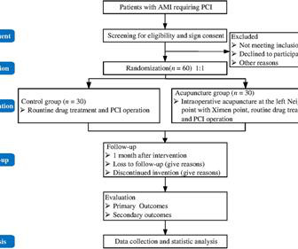
Electroacupuncture (EA) has shown significant efficacy as an adjuvant therapy for many cardiovascular diseases by improving microcirculation and reducing ischemia-reperfusion injury. Currently, effective treatment is not available for SF-NR. However, its effects on SF-NR in the AMI patients during PCI are not clear.
She presented to an outside hospital after several days of malaise and feeling unwell. Application to Today's Case: Today's patient developed ventricular septal rupture the evening after she was admitted to the hospital. At the time of admission, her vital signs were normal. Heart rate was in the 80s.
About 20 minutes later ( on the way to the hospital ) — the patient's CP resolved, and ECG #1 was recorded. That the "culprit" artery spontaneously opened ~20 minutes later while the EMS unit was en route to the hospital ( at which time the patient's CP had resolved — and ECG #1 was obtained ).
My interpretation was: RBBB with hyperacute T-waves in V4-V6 that are all but diagnostic of LAD occlusion vs. post ROSC ischemia. The patient had ROSC and maintained it. A 12-lead ECG was obtained: What do you think? Smith's ECG Blog — Interpretation of a post-resuscitation ECG can be extremely challenging.
The patient was promptly admitted to the hospital for further evaluation. Learning Point: Concordant ST segment elevation can arise from profound ischemia triggered by ventricular tachycardia (VT), or it may represent an exaggerated basal ST change accompanying tachycardia. An initial electrocardiogram (ECG) is provided below.
The differential is: Posterolateral OMI or subendocardial ischemia The distinction between posterior OMI and subendocardial ischemia can be important and sometimes difficult. Ischemic ST depression includes posterior OMI and subendocardial ischemia. Her prior ECG on file is shown below: What are your next steps?
IntroductionDelirium is an acute cognitive or perceptual disturbance that is associated with prolonged hospital and ICU length of stay, therefore, extending recovery time. This is secondary to delayed postoperative cerebral ischemia and infarction caused by vasospasm.7
Background:Remote ischemic conditioning (RIC) with transient cycles of limb ischemia and reperfusion is a safe and promising neuroprotective therapy in acute ischemic stroke (AIS). Stroke, Volume 56, Issue Suppl_1 , Page AWMP9-AWMP9, February 1, 2025. mL (SD 31.18) and Sham 5.83 mL (SD 25.26), P=0.824.
We organize all of the trending information in your field so you don't have to. Join thousands of users and stay up to date on the latest articles your peers are reading.
You know about us, now we want to get to know you!
Let's personalize your content
Let's get even more personalized
We recognize your account from another site in our network, please click 'Send Email' below to continue with verifying your account and setting a password.


Let's personalize your content