This site uses cookies to improve your experience. To help us insure we adhere to various privacy regulations, please select your country/region of residence. If you do not select a country, we will assume you are from the United States. Select your Cookie Settings or view our Privacy Policy and Terms of Use.
Cookie Settings
Cookies and similar technologies are used on this website for proper function of the website, for tracking performance analytics and for marketing purposes. We and some of our third-party providers may use cookie data for various purposes. Please review the cookie settings below and choose your preference.
Used for the proper function of the website
Used for monitoring website traffic and interactions
Cookie Settings
Cookies and similar technologies are used on this website for proper function of the website, for tracking performance analytics and for marketing purposes. We and some of our third-party providers may use cookie data for various purposes. Please review the cookie settings below and choose your preference.
Strictly Necessary: Used for the proper function of the website
Performance/Analytics: Used for monitoring website traffic and interactions
A 60 yo with 2 previous inferior (RCA) STEMIs, stented, called 911 for one hour of chest pain. He had no h/o heartfailure. Here is the presentation ECG for that inferior STEMI: This looks like a large infarct on ECG. Here is the presentation ECG for that inferior STEMI: This looks like a large infarct on ECG.
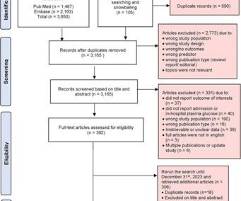
Background Hyperglycemia, characterized by elevated blood glucose levels, is frequently observed in patients with acute coronary syndrome, including ST-elevation myocardial infarction (STEMI). There are conflicting sources regarding the relationship between hyperglycemia and outcomes in STEMI patients. 3.45) and 4.47 (95% CI: 2.54–7.87),
Smith comment 2: I frequently see failure to control BP in patients with acute chest pain or acute heartfailure. The Queen of Hearts once again diagnoses OMI with high confidence: The ED provider recognized the changes in this EKG and called cardiology for a STAT consult.
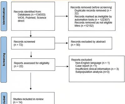
The outcomes of interest were all-cause death and major adverse cardiovascular events (MACE), including acute coronary syndrome (ACS), heartfailure (HF), need for additional revascularization, target vessel revascularization (TVR), SCAD recurrence, and stroke. Approximately 48.5% and 1.3%, respectively.
Here is his ED ECG: There is obvious infero-posterior STEMI. What are you worried about in addition to his STEMI? This was stented. Comments: STEMI with hypokalemia, especially with a long QT, puts the patient at very high risk of Torsades or Ventricular fibrillation (see many references, with abstracts, below).
This is documented as a STEMI in the clinical notes and in the cath report, but certainly does not meet STEMI criteria and is therefore an NSTEMI by definition. For national registry purposes, this will be incorrectly classified as a STEMI.) Most STEMI have peak cTnI greater than 10.0. Large STEMI are approximately 30-80.
This is all but diagnostic of STEMI, probably due to wraparound LAD The cath lab was activated. It was stented. These include: i ) appreciation of how problematic the definition of “acute STEMI” can be; and , ii ) illustration of how dependence on this definition may result in overlooking acute coronary occlusion.
The "criteria" for posterior STEMI are 0.5 There was a normal creatinine and no evidence of heartfailure and no other reason for chronic injury, so it must be acute. Is it STEMI or NonSTEMI? The troponin I returned at 4.1 ng/mL (ULN = 0.030 ng/mL) , diagnostic of myocardial injury. mm STE in one lead. This includes: 1.
He denied any known medical history, specifically: coronary artery disease, hypertension, dyslipidemia, diabetes, heartfailure, myocardial infarction, or any prior PCI/stent. It doesn’t meet any conventional STEMI criteria, but there is patently obvious increased area under the curve. No appreciable skin pallor.
The patient was brought to the ED as a possible Code STEMI and was seen directly by cardiology. Accordingly, in the algorithm by Cai et al for patients with LBBB and ischemic symptoms ( See below ) — the first indication for PCI is clinical: patients with hemodynamic instability or acute heartfailure. So the RCA was stented.
It definitely does not fulfill STEMI criteria, and I would argue that it would not lead to cath lab activation in most centers. In a word — Patient #2 was lucky to have his ECG interpreted by the Queen Of Hearts. As a result — the heart rate of ~115/minute in ECG #1 is a worrisome finding. The ECG shows ST depression in lead V3.
This worried the crew of potential acute coronary syndrome and STEMI was activated pre-hospital. When OMI is captured in this early phase, there exists the highest amount of salvageable myocardium and least likelihood of heartfailure at hospital discharge. A mid-LAD culprit lesion was identified and stented.
The patient was then taken to the cath lab an found to have a proximal RCA 100% thrombotic occlusion which was successfully stented. The patient was then taken to the cath lab an found to have a proximal RCA 100% thrombotic occlusion which was successfully stented. Progression of V2 showing posterior involvement.
Case submitted by Andrew Grimes, Advanced Care paramedic, with additions from Jesse McLaren and Smith An 84-year-old male with a notable cardiac history (CABG, multiple stents) woke at 0500hrs with pressure in his chest, diaphoresis, and light-headedness. STEMI criteria are only 43% sensitive for OMI.
Unfortunately, the ECG was interpreted as no significant change from prior , "no STEMI"!! Approximately 5 minutes after ROSC, this ECG was obtained (about 45 minutes after arrival): Obvious anterolateral OMI, and STEMI criteria positive for those who care or need it. He was sent back to the waiting room, where he suffered a VF arrest.
After stent deployment, we often see improvement in the ST-T within seconds or minutes. Here is the final angiogram following placement of a stent in the ostial RCA. 2:04 PM, post stent deployment You can see that even after complete restoration of flow, the ECG still looks terrible, V most of all.
We organize all of the trending information in your field so you don't have to. Join thousands of users and stay up to date on the latest articles your peers are reading.
You know about us, now we want to get to know you!
Let's personalize your content
Let's get even more personalized
We recognize your account from another site in our network, please click 'Send Email' below to continue with verifying your account and setting a password.


Let's personalize your content