This site uses cookies to improve your experience. To help us insure we adhere to various privacy regulations, please select your country/region of residence. If you do not select a country, we will assume you are from the United States. Select your Cookie Settings or view our Privacy Policy and Terms of Use.
Cookie Settings
Cookies and similar technologies are used on this website for proper function of the website, for tracking performance analytics and for marketing purposes. We and some of our third-party providers may use cookie data for various purposes. Please review the cookie settings below and choose your preference.
Used for the proper function of the website
Used for monitoring website traffic and interactions
Cookie Settings
Cookies and similar technologies are used on this website for proper function of the website, for tracking performance analytics and for marketing purposes. We and some of our third-party providers may use cookie data for various purposes. Please review the cookie settings below and choose your preference.
Strictly Necessary: Used for the proper function of the website
Performance/Analytics: Used for monitoring website traffic and interactions
a developer of cellular and cell-derived therapeutics for the treatment of cardiovascular and pulmonary diseases, today announced the primary endpoint results of the open label roll-in cohort of the CardiAMP Cell Therapy in Chronic Myocardial Ischemia Trial. Getty Images milla1cf Thu, 05/02/2024 - 10:12 May 2, 2024 — BioCardia, Inc. ,
Beyond its role in circadian rhythm regulation, REV-ERB could significantly influence physiological and pathological processes related to cardiovascular health, including atherosclerosis, myocardial ischemia/reperfusion injury, and heartfailure.
This EKG is diagnostic of transmural ischemia of the inferior wall. If it is angina, lowering the BP with IV Nitroglycerine may completely alleviate the pain and the (unseen) ECG ischemia. Smith comment 2: I frequently see failure to control BP in patients with acute chest pain or acute heartfailure.
Myocardial ischemia may induce myocardial fibrosis, a condition that progressively leads to ventricular remodeling, heightening the risk of heartfailure. 68 Ga-FAPI-04 PET/CT shows promise in assessing fibroblast activation in patients with early myocardial infarction characterized by prolonged myocardial ischemia.
It should be kept in mind that on occasions, beta-one agonist can result in increased ventricular ectopy e.g., in severe myocardial ischemia (by increasing myocardial demand), or sometimes with congenital long-QT syndrome. Smith, this can be accomplished by either using beta-one agonists or temporary transvenous pacing.
Additionally, ischemia-reperfusion injury following cardioplegic arr. Cardiopulmonary bypass induces a systemic inflammatory response and alterations in fluid homeostasis, resulting in generalized tissue edema.
DISCUSSION: The 12-lead EKG EMS initially obtained for this patient showed severe ischemia, with profound "infero-lateral" ST depression and reciprocal ST elevation in lead aVR. The ECG cannot diagnose the etiology of ischemia; it only the presence of ischemia, from whatever etiology.
Journal of the American Heart Association, Ahead of Print. BackgroundThe early assessment of heartfailure (HF) risk in patients with acute coronary syndrome (ACS) can help reduce mortality.
Are you confident there is no ischemia? The heart rate is about 130 bpm. The heart rate could be compatible with that of a 2:1 conducted atrial flutter. Primary VT , and the VT with tachycardia is causing ischemia with chest discomfort (supply-demand mismatch/type 2 MI)? Do you agree with this strategy?
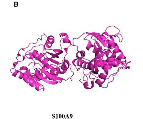
This article evaluates the utility of S100A8/A9 protein as a biomarker and therapeutic target for diagnosing cardiovascular diseases, considering its structural features, fundamental biological properties, and its multifaceted influence on cardiovascular conditions including atherosclerosis, myocardial infarction, myocardial ischemia/reperfusion injury, (..)
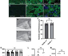
Myocardial damage caused, for example, by cardiac ischemia leads to ventricular volume overload resulting in increased stretch of the remaining myocardium. Conversely, in response to extensive myocardial damage, cardiomyocytes in the adult zebrafish heart and neonatal mice proliferate and completely regenerate the damaged myocardium.
In any case, the ECG is diagnostic of severe ischemia and probably OMI. So this could be myocarditis but in my opinion needs an angiogram before making that diagnosis. == Dr. Nossen Comment/Interpretation: Evaluation of ischemia on an ECG can be very challenging. Concordant STE of 1 mm in just one lead or 2a.
The patient has heartfailure as a result of this event. If this STD were due to LVH or to subendocardial ischemia, rather than posterior OMI, it would be maximal in V5 and V6. How could an occlusion (Occlusion MI, OMI) that results in the loss of a massive amount of myocardium and results in HeartFailure be missed?
In the heart, autophagy is regulated mainly through mitophagy due to the metabolic changes of cardiomyocytes caused by ischemia and hypoxia. Myocardial remodeling is characterized by gradual heart enlargement, cardiac dysfunction, and extraordinary molecular changes.
His medical history includes hypertension, a decade-long battle with diabetes, ischemic heart disease, a coronary bypass graft surgery ten years ago, a diagnosis of congestive heartfailure for the last five years, and a prior ICD implantation five years ago. Thus VT is very probable.
NOTE: It's important to correlate ongoing circumstances at the time that a prior tracing was done ( ie, Was the patient stable and asymptomatic — or were they having chest pain, an exacerbation of heartfailure, or some other ongoing process at the time the prior ECG was recorded? ). Cardiol 27:674-677, 2004 ).
ET Murphy Ballroom 4 Comparison of an "Inclisiran First" Strategy with Usual Care in Patients With Atherosclerotic Cardiovascular Disease: Results From the VICTORION-INITIATE Randomized Trial Targeting Weight Loss to Personalize the Prevention of Type 2 Diabetes Once-weekly Semaglutide in Patients with HeartFailure With Preserved Ejection Fraction, (..)
Most such rhythms in the setting of ischemia are VF and will not convert without defibrillation. NT-pro-BNP peaked at 4831, consistent with heartfailure. Instead, antiarrhythmic drugs such as amiodarone or ß-blockers may be needed — and/or treatment targeted to correcting ischemia. Acute ischemia?
The fact that R waves 2 through 6 are junctional does make ischemia more difficult to interpret -- but not impossible. The Queen of Hearts does not care about rhythm analysis, she simply looks at the ECG and decides whether it represents OMI or not.
BACKGROUND:Cardiolipin is a mitochondrial-specific phospholipid that maintains integrity of the electron transport chain (ETC) and plays a central role in myocardial ischemia/reperfusion injury. Circulation, Ahead of Print. Tafazzin is an enzyme that is required for cardiolipin maturation.
Analyses of cardiovascular magnetic resonance imaging (MRI) in the MESA cohort have shown that LV sphericity is an important mode of variation in cardiac structure that is associated with ischemia, heartfailure, and atrial fibrillation.
Accordingly, in the algorithm by Cai et al for patients with LBBB and ischemic symptoms ( See below ) — the first indication for PCI is clinical: patients with hemodynamic instability or acute heartfailure. So there is now high pre-test probability + refractory ischemia + Modified Sgarbossa + dynamic ECG changes.
IntroductionTransient Ischemic Attack (TIA) is a common neurologic condition characterized by temporary, focal cerebral ischemia that results in reversible neurological deficits without tissue infarction. Stroke: Vascular and Interventional Neurology, Volume 3, Issue S2 , November 1, 2023.
There is broad subendocardial ischemia as demonstrated by STE aVR with concomitant STD that almost appears appropriately maximal in Leads II and V5. There is LBBB-like morphology with persistent patterns of subendocardial ischemia. This is the initial ECG: The QRS is widened with a regular cadence, and there are no discernable P waves.
They have heartfailure sometimes. That is, there is renal ischemia due to clogging of capillaries, which leads to increased erythropoietin secretion and this causes decompensated erythrocytosis. So they do badly, but it develops only very late and in very few also. It produces cardiomegaly on X-ray in ASD Eisenmenger.
Diffuse ST depression with ST elevation in aVR: Is this pattern specific for global ischemia due to left main coronary artery disease? Ischemia b. ST depression: is it ischemia? Does this patient have hypertension and/or heartfailure that has worsened? Reference: Knotts RJ , Wilson JM, Kim E, Huang HD, Birnbaum Y.
There was no evidence of ischemia. In addition to ruling out rate-slowing medication serum electrolyte disorders and/or ischemia/infarction as potential causes of bradyarrhythmias one should also rule out hypothyroidism + sleep apnea. We are not told how ischemia has been ruled out in this case. Hyperkalemia.
In the ISCHEMIA (International Study of Comparative Health Effectiveness with Medical and Invasive Approaches) trial, researchers examined the risk of ischemic events in patients with stable coronary artery disease. years, with 57.1% occurring within 30 days after CABG. Original article: Redfors B et al.
All of this appears to be consistent with "No Reflow", or small vessel occlusion with persistent ischemia in spite of an open artery. --There is persistent ST elevation in leads V1-V4, with a lot of STE in V4 (another bad sign). The best predictor of good blush and high TMP is the ECG, specifically resolution of ST Elevation on the ECG.
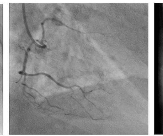
100% occluded RCA with TIMI 0 flow Post drug-eluting stent placement with TIMI 3 flow While in the cath lab, she transiently developed complete heart block and became hypotensive requiring transvenous pacemaker placement and transient pressors. Peak troponin T was 3.00 ng/mL (highly elevated). Post-cath ECG with resolution of acute changes.
These include: i ) Use of rate-slowing medication ( ie, ß-blockers, digoxin, verapamil/diltiazem, etc. ) ; ii ) Acute or recent infarction or ischemia; iii ) Hypothyroidism; iv ) Neurologic injury; v ) Electrolyte disturbance; and , vi ) Sleep apnea.
It is important to appreciate that even before looking at the ECG itself — the chance of true chamber enlargement is greatly increased IF the patient is older, and has longstanding hypertension and/or underlying heart disease likely to predispose to chamber enlargement (ie, heartfailure, cardiomyopathy, valvular disease, etc. ).
The flutter waves can conceal or mimic ischemic repolarization findings, but here I don't see any obvious findings of OMI or subendocardial ischemia. The rhythm is 2:1 atrial flutter. The QRS is around 100 msec wide (narrow), but with very abnormal morphology including a large R-wave in V1, deep S-wave in I, R-wave in aVR.
At present, the main methods to treat ischemic heart disease are drug therapy, intervention and operation. These methods only alleviate symptoms of heartfailure and myocardial ischemia and improve patients' quality of life by partially restoring myocardial reperfusion.
Dilation: The chambers of the heart expand, making the walls thinner. Both forms of enlargement may compromise the heart’s ability to pump blood efficiently, leading to further complications like heartfailure. What Causes an Enlarged Heart? Here are some of the most common causes: 1.
There is no evidence of infarction or ischemia. NT-proBNP values less than 300 pg/ml have a 99% negative predictive value for excluding congestive heartfailure. A cutoff of 1200 pg/ml for patients with a normal eGFR is very specific for heartfailure. Troponin I was 0.054 ng/mL NT-ProBNP was 8316 (0-900 pg/mL). "
Central illustration: In those presenting With monomorphic VT, undergoing coronary ischemia assessment was associated With improved 12-month event-free survival from the primary endpoint of VT recurrence, ICD therapy, heartfailure hospitalization, and death.
The first task when assessing a wide complex QRS for ischemia is to identify the end of the QRS. The ST segment changes are compatible with severe subendocardial ischemia which can be caused by type I MI from ACS or potentially from type II MI (non-obstructive coronary artery disease with supply/demand mismatch). What do you think?
Background The clinical presentation of left ventricular free wall rupture (LVFWR) varies ranging from uneventful condition to congestive heartfailure. Case summary Here we report two cases of LVFWR with different clinical presentation and notable outcome.
Critical limb-threatening ischemia (CLTI) represents the end stage of peripheral artery disease (PAD), when poor circulation due to blockages in the arteries causes symptoms including numbness, sores that will not heal, gangrene, and extreme pain. When we save a limb, we save a life.
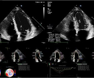
Risk stratification of cardiovascular death and treatment strategies in patients with heartfailure (HF), the optimal timing for valve replacement, and the selection of patients for implantable cardioverter defibrillators are based on an echocardiographic calculation of left ventricular ejection fraction (LVEF) in most guidelines.
We organize all of the trending information in your field so you don't have to. Join thousands of users and stay up to date on the latest articles your peers are reading.
You know about us, now we want to get to know you!
Let's personalize your content
Let's get even more personalized
We recognize your account from another site in our network, please click 'Send Email' below to continue with verifying your account and setting a password.


Let's personalize your content