This site uses cookies to improve your experience. To help us insure we adhere to various privacy regulations, please select your country/region of residence. If you do not select a country, we will assume you are from the United States. Select your Cookie Settings or view our Privacy Policy and Terms of Use.
Cookie Settings
Cookies and similar technologies are used on this website for proper function of the website, for tracking performance analytics and for marketing purposes. We and some of our third-party providers may use cookie data for various purposes. Please review the cookie settings below and choose your preference.
Used for the proper function of the website
Used for monitoring website traffic and interactions
Cookie Settings
Cookies and similar technologies are used on this website for proper function of the website, for tracking performance analytics and for marketing purposes. We and some of our third-party providers may use cookie data for various purposes. Please review the cookie settings below and choose your preference.
Strictly Necessary: Used for the proper function of the website
Performance/Analytics: Used for monitoring website traffic and interactions
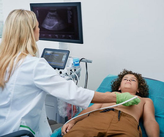
Interpreting cardiac ultrasounds has become more complex and time-consuming for clinicians: With the shift toward a more quantitative analysis, there are more parameters for clinicians to measure. Heres a look at how AI ultrasound and other tech innovations are helping cardiologists work faster while driving better outcomes for patients.
Scientists have found soundwaves from low-intensity focused ultrasound aimed at a place deep in the brain called the insula can reduce both the perception of pain and other effects of pain, such as heart rate changes.
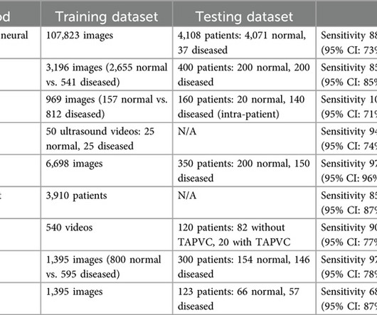
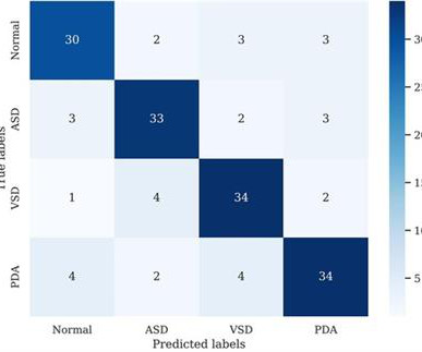
BackgroundCongenital heartdisease (CHD) is a major contributor to morbidity and infant mortality and imposes the highest burden on global healthcare costs. Most studies utilized deep learning models using either ultrasound or echocardiographic images.
Cardiovascular ultrasound has played a key role in the evolution of early diagnosis of structural heartdisease, led by a technology pioneered by Philips: the ‘transesophageal echocardiography’ (TEE) ultrasound transducer. In structural heartdisease, the quality of a 3D TEE image can help save lives.
Speed of ultrasound in body tissues is around: A. 1500 meters per second Speed of ultrasound is almost that in water, though there is some difference between various tissues. Reflections of ultrasound occur at interfaces between tissues and between blood and tissues, due to the difference in velocity. 20,000 meters per second B.
Food and Drug Adminstration (FDA) has approved DEFINITY (Perflutren Lipid Microsphere) as an ultrasound enhancing agent for use in pediatric patients with suboptimal echocardiograms, including those who have undergone heart transplant, or have Kawasaki disease or a congenital cardiovascular anomaly.
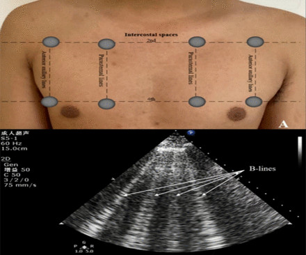
Objectives Prognostic impact of lung ultrasound-derived B-lines (LUS-BL) in heart failure with mildly reduced left ventricular ejection fraction (HFmrEF) patients remains elusive. We evaluated the correlation between LUS-BL and prognosis in HFmrEF patients.
These features can help physicians perform cardiac procedures more efficiently in the areas of diagnostics, structural heartdisease, and electrophysiology. Available exclusively with the ACUSON Origin, the new AcuNav Lumos 4D ICE (intracardiac echocardiography) catheter provides advanced imaging for complex heart procedures.
milla1cf Wed, 06/05/2024 - 20:37 June 5, 2024 — UltraSight , a pioneer in digital health transforming cardiac imaging with artificial intelligence, is collaborating with Mayo Clinic on a new endeavor with the goal of enhancing cardiac care by harnessing the power of AI in point-of-care ultrasound.
Zeke and Zane were both diagnosed with hypoplastic left heart syndrome (HLHS) before birth. As far as congenital heartdiseases go, HLHS falls on the rarer end of the spectrum. The Heart Institute collaborates with the CFCC through the Fetal Cardiology Program.
Ultrasound techniques currently used in echocardiography uses frame rates from 30-150 frames/s. This limits its temporal resolution for very short lived events, especially in pediatric and congenital heartdisease with faster heart rates compared to adults [1]. J Am Coll Cardiol. 2024 Jan 2;83(1):63-81. 2023.10.025.
Paul Dudley White, the famous physician who has taught many a luminary in the field of cardiology once wrote that heartdisease before eighty is our fault and not God’s will or nature’s will. This means that he recognized long back, the role of life-style modification in preventing heartdisease.
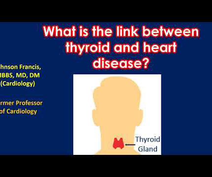
Heartdisease can occur with both increased function of the thyroid gland and decreased function of the thyroid gland. When thyroid function is increased, heart rate increases and the work load of the heart increases. In severe cases heart failure may occur.
Congenital heartdisease is a prevalent birth defect, accounting for approximately one-third of major birth defects. However, the emergence of deep learning in computer vision has paved the way for detecting subtle changes in chest x-rays, such as lung vessel density, enabling the detection of congenital heartdisease in children.
Cardiac device therapy is frequently required for individuals with adult congenital heartdisease (ACHD), whether it be for bradyarrhythmia, ventricular tachyarrhythmia, or cardiac resynchronization therapy (CRT).
This year’s program, “Global Echocardiography: Innovations in Diagnosis and Beyond,” is described as an educational experience gathering global experts and enthusiasts in echocardiography, hosted by the largest global organization for cardiovascular ultrasound imaging serving physicians, sonographers, nurses, and scientists.
Food and Drug Administration (FDA) has granted 510(k) clearance for its first-of-a-kind, AI-powered AISAP CARDIO point-of-care ultrasound (POCUS) software platform. We know that structural heartdisease and heart failure are the leading causes of hospitalization and morbidity in the U.S.
BackgroundIschemic cardiomyopathy (ICM) is the end stage of ischemic heartdisease, in which ventricular remodeling contributes to a fatal ventricular arrhythmia, worsens heart function and unfavorable outcomes, and is related to persistent chronic inflammation.
Echocardiography remains the reference-standard imaging technique for assessing valvular heartdisease (VHD), but artifacts like the ‘color Doppler stripe’ can complicate diagnosis. This artifact is not widely.
It’s a celebration of the amazing innovation that has occurred in structural heartdisease therapy over the last 20 years and I’m proud to be part of it. He also played a key role in developing intravascular ultrasound, as well as the U.S. standard-of-care for managing postpartum hemorrhage, the JADA System.
Carcinoid heartdisease (CHD) caused by neuroendocrine tumours (NET) is associated with an increased morbidity and mortality due to valvular dysfunction and right sided heart failure. The present study aimed t.
He arrived in the ED and had an immediate bedside cardiac ultrasound while this ECG was being recorded. The bedside ultrasound (video not available) reportedly showed only a slightly reduced LV function. A regular wide complex tachycardia in a young patient with no history of heartdisease is very likely to be AVRT.
Purpose This study aims to evaluate deep learning (DL) denoising reconstructions for image quality improvement of Doppler ultrasound (DUS)-gated fetal cardiac MRI in congenital heartdisease (CHD). Cine imaging was acquired using a balanced steady-state free precession (bSSFP) sequence with Doppler ultrasound gating.
A large randomized trial showed no significant differences to demonstrate that using artificial intelligence (AI) to aid clinical decision-making in assessing heartultrasounds is as effective as current practice at identifying all-comers with suspected heartdisease who may benefit from invasive investigation and treatment.
Fetal cardiac intervention team included pediatric cardiology imaging specialist, fetal cardiology nurse practitioner, two interventional pediatric cardiologists, ultrasound radiologist, maternal-fetal medicine physician, fetal anaesthesiologist and maternal anaesthesiologist. Procedure is done under high quality ultrasound imaging guidance.
24: Joint American College of Cardiology/Journal of the American College of Cardiology Late-Breaking Clinical Trials (Session 402) Saturday, April 6 9:30 – 10:30 a.m.
We describe a fetus with prenatal echocardiographic findings of BDA and right aortic arch mirror-image branching (RAA-MIB) combined with congenital heartdisease. Prenatal ultrasound diagnosis of BDA is important and requires a combination of 2D grayscale, CDFI, and STIC images to assist in scanning.
Schedule Regular Checkups With Your Cardiologist: Early detection and management of heart conditions are crucial for maintaining good health. Electrophysiology: This specialized area focuses on heart rhythm problems, like atrial fibrillation (AFib).
A bedside cardiac ultrasound was normal. The P wave is positive in lead aVL of ECG #3, which means it is a low atrial (or probably coronary sinus) rhythm — which of itself is not necessarily “abnormal” in a child if there is no other sign of underlying heartdisease. His chest was tender. He wrote: "ECG 1 - shows wide ???IVCD
Introduction:Dextrocardia is a rare congenital condition where the heart's apex points to the right, with an incidence of about 0.01%. Patients usually have a normal life expectancy unless other structural heartdiseases are present. An intravascular ultrasound was also performed, which was negative for vessel dissection.
Schedule Regular Checkups With Your Cardiologist: Early detection and management of heart conditions are crucial for maintaining good health. Electrophysiology: This specialized area focuses on heart rhythm problems, like atrial fibrillation (AFib).
For example, by integrating Ventripoint’s AI-powered heart-scanning technology, which turns ultrasound images of the heart into MRI-quality heart images, InView provides pediatric cardiologists with access to MRI-quality heart images at a fraction of the cost and time needed for traditional MRIs.
So today i wanted to talk to you about what each heart test tells us about these different aspects of heartdisease Tests that tell you about the heart as a pump The most commonly used test to assess the heart as a pump is an echocardiogram. If the heart has been left damaged, then that part of.
Identify accumulated blood clots that can result in heartdiseases, cancer, or emphysema. Magnetic Resonance-Guided Ultrasound It is an MRI-based therapeutic technique that makes use of ultrasonic pulses to remove the target tissue. The procedure is simple, painless, and takes significantly less time to conduct.
Smith comment: This patient did not have a bedside ultrasound. Had one been done, it would have shown a feature that is apparent on this ultrasound (however, this patient's LV function would not be as good as in this clip): This is recorded with the LV on the right. In fact, bedside ultrasound might even find severe aortic stenosis.
Image courtesy: Philips christine.book Wed, 06/12/2024 - 14:07 June 12, 2024 — Royal Philips has announced its next-generation AI-enabled cardiovascular ultrasound platform to help speed up cardiac ultrasound analysis with proven AI technology and reduce the burden on echocardiography labs.
Her bedside cardiac ultrasound was normal We decided to cardiovert her since the time of onset was very recent. In the study below, almost all patients had serious heartdisease and they are less likely to convert with electricity alone. But when you see this, you should suspect that the AV node is not well.
A bedside cardiac ultrasound was performed with a parasternal long axis view demonstrated below: There is a large pericardial effusion with collapse of the right ventricle during systole. The beat-to-beat variation in QRS amplitude and morphology is electrical alternans. This patient is only pseudo-stable. She has already had syncope.
During echocardiography, a transducer transmits the ultrasound beam towards the heart. Echoes received by the transducer from various structures of the heart are analysed by the echocardiograph and a graphical representation displayed on the monitor. This view images the heart from the base to apex long axis view.
Getty Images milla1cf Mon, 04/01/2024 - 08:21 April 1, 2024 — Roughly 25,000 Americans die each year from valvular heartdisease, but researchers from Rutgers Health and other institutions conclude that new technology could soon help doctors slash that number. “We What is Valvular HeartDisease?
J Am Heart Assoc. PMID: 34775811; PMCID: PMC9075358 A bedside ultrasound was performed, shown here: Parasternal short axis view demonstrating inferior LV wall motion akinesis Apical 2 chamber view again demonstrating inferior LV wall akinesis The cath lab was not activated based on the ECG and bedside echo. 2021 Dec 7;10(23):e022866.
Now, with Caption AI technology, clinicians using Vscan Air SL handheld ultrasound will have access to real-time, step-by-step guidance to capture diagnostic-quality images and automated ejection fraction estimation to help inform clinical decisions across cardiac settings. Strom , M.D.,
Getty Images milla1cf Tue, 01/16/2024 - 14:11 January 16, 2024 — Artificial intelligence (AI) has the potential to detect rheumatic heartdisease (RHD) with the same accuracy as a cardiologist, according to new research demonstrating how sophisticated deep learning technology can be applied to this disease of inequity.
We organize all of the trending information in your field so you don't have to. Join thousands of users and stay up to date on the latest articles your peers are reading.
You know about us, now we want to get to know you!
Let's personalize your content
Let's get even more personalized
We recognize your account from another site in our network, please click 'Send Email' below to continue with verifying your account and setting a password.


Let's personalize your content