This site uses cookies to improve your experience. To help us insure we adhere to various privacy regulations, please select your country/region of residence. If you do not select a country, we will assume you are from the United States. Select your Cookie Settings or view our Privacy Policy and Terms of Use.
Cookie Settings
Cookies and similar technologies are used on this website for proper function of the website, for tracking performance analytics and for marketing purposes. We and some of our third-party providers may use cookie data for various purposes. Please review the cookie settings below and choose your preference.
Used for the proper function of the website
Used for monitoring website traffic and interactions
Cookie Settings
Cookies and similar technologies are used on this website for proper function of the website, for tracking performance analytics and for marketing purposes. We and some of our third-party providers may use cookie data for various purposes. Please review the cookie settings below and choose your preference.
Strictly Necessary: Used for the proper function of the website
Performance/Analytics: Used for monitoring website traffic and interactions
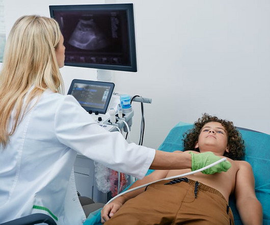
Food and Drug Adminstration (FDA) has approved DEFINITY (Perflutren Lipid Microsphere) as an ultrasound enhancing agent for use in pediatric patients with suboptimal echocardiograms, including those who have undergone heart transplant, or have Kawasaki disease or a congenital cardiovascular anomaly.
Cardiovascular ultrasound has played a key role in the evolution of early diagnosis of structural heartdisease, led by a technology pioneered by Philips: the ‘transesophageal echocardiography’ (TEE) ultrasound transducer. In structural heartdisease, the quality of a 3D TEE image can help save lives.
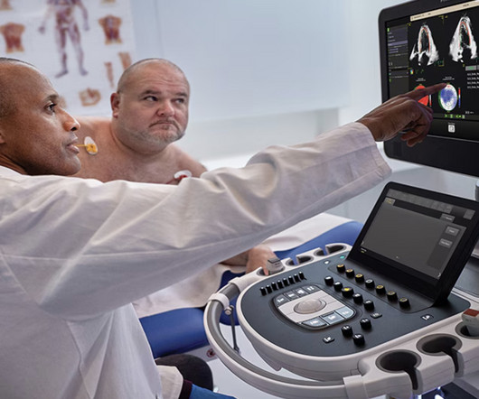
Image courtesy: Philips christine.book Wed, 06/12/2024 - 14:07 June 12, 2024 — Royal Philips has announced its next-generation AI-enabled cardiovascular ultrasound platform to help speed up cardiac ultrasound analysis with proven AI technology and reduce the burden on echocardiography labs.
Getty Images milla1cf Tue, 01/16/2024 - 14:11 January 16, 2024 — Artificial intelligence (AI) has the potential to detect rheumatic heartdisease (RHD) with the same accuracy as a cardiologist, according to new research demonstrating how sophisticated deep learning technology can be applied to this disease of inequity.
Zeke and Zane were both diagnosed with hypoplastic left heart syndrome (HLHS) before birth. As far as congenital heartdiseases go, HLHS falls on the rarer end of the spectrum. The Heart Institute collaborates with the CFCC through the Fetal Cardiology Program.
Ultrasound techniques currently used in echocardiography uses frame rates from 30-150 frames/s. This limits its temporal resolution for very short lived events, especially in pediatric and congenital heartdisease with faster heart rates compared to adults [1]. J Am Coll Cardiol. 2024 Jan 2;83(1):63-81.
This year’s program, “Global Echocardiography: Innovations in Diagnosis and Beyond,” is described as an educational experience gathering global experts and enthusiasts in echocardiography, hosted by the largest global organization for cardiovascular ultrasound imaging serving physicians, sonographers, nurses, and scientists.
Background Axillary arterial access (AAA) in pediatricheart catheterizations is undervalued. Methods We retrospectively reviewed children with congenital heartdiseases (CHDs) who received trans-axillary arterial catheterizations between January 2019 and February 2023.
Fetal cardiac intervention team included pediatric cardiology imaging specialist, fetal cardiology nurse practitioner, two interventional pediatric cardiologists, ultrasound radiologist, maternal-fetal medicine physician, fetal anaesthesiologist and maternal anaesthesiologist. Reference Ryan Callahan, Kevin G. Esch, Lynn A.
For example, by integrating Ventripoint’s AI-powered heart-scanning technology, which turns ultrasound images of the heart into MRI-quality heart images, InView provides pediatric cardiologists with access to MRI-quality heart images at a fraction of the cost and time needed for traditional MRIs.
A bedside cardiac ultrasound was normal. The P wave is positive in lead aVL of ECG #3, which means it is a low atrial (or probably coronary sinus) rhythm — which of itself is not necessarily “abnormal” in a child if there is no other sign of underlying heartdisease. His chest was tender. He wrote: "ECG 1 - shows wide ???IVCD
During echocardiography, a transducer transmits the ultrasound beam towards the heart. Echoes received by the transducer from various structures of the heart are analysed by the echocardiograph and a graphical representation displayed on the monitor. This view images the heart from the base to apex long axis view.
We organize all of the trending information in your field so you don't have to. Join thousands of users and stay up to date on the latest articles your peers are reading.
You know about us, now we want to get to know you!
Let's personalize your content
Let's get even more personalized
We recognize your account from another site in our network, please click 'Send Email' below to continue with verifying your account and setting a password.


Let's personalize your content