This site uses cookies to improve your experience. To help us insure we adhere to various privacy regulations, please select your country/region of residence. If you do not select a country, we will assume you are from the United States. Select your Cookie Settings or view our Privacy Policy and Terms of Use.
Cookie Settings
Cookies and similar technologies are used on this website for proper function of the website, for tracking performance analytics and for marketing purposes. We and some of our third-party providers may use cookie data for various purposes. Please review the cookie settings below and choose your preference.
Used for the proper function of the website
Used for monitoring website traffic and interactions
Cookie Settings
Cookies and similar technologies are used on this website for proper function of the website, for tracking performance analytics and for marketing purposes. We and some of our third-party providers may use cookie data for various purposes. Please review the cookie settings below and choose your preference.
Strictly Necessary: Used for the proper function of the website
Performance/Analytics: Used for monitoring website traffic and interactions
Perimembranous VSD is the commonest type of ventricular septal defect noted in children. Large VSDs can be associated with heart failure in infancy and may need surgery. Device closure is not that well established in perimembranous VSD as in case of muscular VSD.
Diagrammatic representation of VSD Eisenmenger. The other Eisenmenger syndromes are not called Eisenmenger complex, only VSD Eisenmenger is called Eisenmenger complex. Large ASDs usually develop Eisenmenger syndrome, may be after decades, not like early development of Eisenmenger syndrome in VSD and PDA.
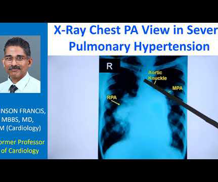
In VSD Eisenmenger and PDA Eisenmenger, you would not expect right atrial enlargement. In VSD and PDA Eisenmenger, the heart size decreases when pulmonary hypertension develops, due to decrease in the shunt. In ASD, shunt is decreasing in Eisenmenger but right atrial enlargement is a component for cardiomegaly.
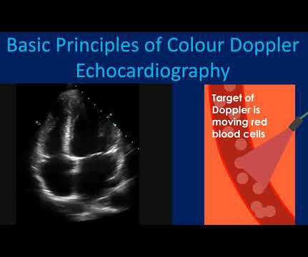
For example, when the VSD is small, the pressure difference between the right and left ventricles is high. Colour Doppler echocardiography is very useful in giving a quick visual assessment of regurgitation and stenosis of heart valves. It will also show abnormal flows as in an atrial or ventricular septal defect.
The VSD is partly overrided by the aorta. I am sure that most of you are familiar with echocardiography would have come at the diagnosis and differential diagnosis by this time because it is a very simple view. And this is a better annotated view and I am showing the ventricular septal defect here. Right ventricle, left ventricle.
But in a VSD with pulmonary hypertension A wave is not prominent. It will not occur in the presence of a large VSD which equalizes both right ventricular and left ventricular pressures. Right atrial hypertrophy as in tricuspid stenosis, pulmonary stenosis and pulmonary hypertension.
We organize all of the trending information in your field so you don't have to. Join thousands of users and stay up to date on the latest articles your peers are reading.
You know about us, now we want to get to know you!
Let's personalize your content
Let's get even more personalized
We recognize your account from another site in our network, please click 'Send Email' below to continue with verifying your account and setting a password.


Let's personalize your content