This site uses cookies to improve your experience. To help us insure we adhere to various privacy regulations, please select your country/region of residence. If you do not select a country, we will assume you are from the United States. Select your Cookie Settings or view our Privacy Policy and Terms of Use.
Cookie Settings
Cookies and similar technologies are used on this website for proper function of the website, for tracking performance analytics and for marketing purposes. We and some of our third-party providers may use cookie data for various purposes. Please review the cookie settings below and choose your preference.
Used for the proper function of the website
Used for monitoring website traffic and interactions
Cookie Settings
Cookies and similar technologies are used on this website for proper function of the website, for tracking performance analytics and for marketing purposes. We and some of our third-party providers may use cookie data for various purposes. Please review the cookie settings below and choose your preference.
Strictly Necessary: Used for the proper function of the website
Performance/Analytics: Used for monitoring website traffic and interactions
Speed of ultrasound in body tissues is around: A. 1500 meters per second Speed of ultrasound is almost that in water, though there is some difference between various tissues. Reflections of ultrasound occur at interfaces between tissues and between blood and tissues, due to the difference in velocity. 20,000 meters per second B.
Coronary Intravascular Ultrasound (IVUS) equipment consists of an IVUS catheter, pullback device and the imaging console. Incomplete stent apposition can be detected by intravascular ultrasound. If lesion lengths have to be assessed, motorized pullback is required. For assessing lesion morphology a manual pullback can also be done.
In harmonic imaging, if the frequency of ultrasound transmitted is 2.5 MHz, reception is on: A. 7 MHz Correct answer: B. 5 MHz If the transmit and receive frequency are the same in echocardiography, the image quality may be poorer because of higher levels interference and artifacts.
The ultrasound reflected from red blood cells moving away from the transducer has: A. Lower frequency and colour coded blue Ultrasound reflected from red blood cells moving towards the transducer will be perceived as having higher frequency. Higher frequency and is colour coded red B. Higher frequency and colour coded blue C.
The principle of Doppler is that the frequency of sound wave coming from an object which is moving towards the ultrasound probe increases while that from an object moving away from the probe decreases. Colour Doppler echocardiography receives the ultrasound signals reflected from moving red blood cells in the heart.
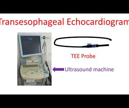
Echocardiogram is an image of the heart using ultrasound. An ultrasound beam is transmitted into the body using a device known as transducer. Esophagus or food pipe is just behind the heart so that the distance which the ultrasound beam has to travel to reach the heart is also small.
Fetal cardiac intervention team included pediatric cardiology imaging specialist, fetal cardiology nurse practitioner, two interventional pediatric cardiologists, ultrasound radiologist, maternal-fetal medicine physician, fetal anaesthesiologist and maternal anaesthesiologist. Reference Ryan Callahan, Kevin G. Esch, Lynn A.
Ultrasound image of the heart – echocardiogram, showing fluid collection around the heart, marked as PE, short for pericardial effusion. Collection of fluid within the covering of the heart is called pericardial effusion. If it is severe enough to compress the heart, it prevents proper filling of the heart and blood pressure falls.
Plaque regression can be demonstrated by ultrasound evaluation of the carotids which are easily accessible. A decrease in fat deposits can reverse the process of atherosclerotic cardiovascular disease, if meticulously followed over a long period. Regular exercise can bring down the blood pressure in the long run.
J Cardiovasc Ultrasound. J Cardiovasc Ultrasound. Ha J et al. Therapeutic strategies for diastolic dysfunction: a clinical perspective. 2009 Sep;17(3):86-95. Park JH et al. Use and Limitations of E/e’ to Assess Left Ventricular Filling Pressure by Echocardiography. 2011 Dec;19(4):169-73. Møller JE et al.
Pericardial effusion is usually confirmed by an echocardiogram (ultrasound study of the heart). When the quantity is large enough to compress the heart, the person may feel breathless or dizzy because of a fall in blood pressure. Sometimes mild pericardial effusion may be detected by an echocardiogram done for other causes.
During echocardiography, a transducer transmits the ultrasound beam towards the heart. It is used in the emergency department, at bedside, in the intensive care unit as well as in the operating room. Hence a basic knowledge is needed for all physicians and paramedics.
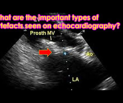
Lower energy side beams of ultrasound known as sidelobes can reflect off lateral structures and get mapped to the central image, causing sidelobe artefacts. Multiple reflections from strong reflectors of ultrasound like pericardium and pleura can cause mirror images of structures being imaged.
Doppler echocardiography is best done with the ultrasound beam: A. Parallel to the direction of blood flow B. Perpendicular to the direction of blood flow C. At an angle of 45 degrees to the direction of blood flow D. At an angle of 135 degrees to the direction of blood flow Correct answer: A.
We organize all of the trending information in your field so you don't have to. Join thousands of users and stay up to date on the latest articles your peers are reading.
You know about us, now we want to get to know you!
Let's personalize your content
Let's get even more personalized
We recognize your account from another site in our network, please click 'Send Email' below to continue with verifying your account and setting a password.


Let's personalize your content