This site uses cookies to improve your experience. To help us insure we adhere to various privacy regulations, please select your country/region of residence. If you do not select a country, we will assume you are from the United States. Select your Cookie Settings or view our Privacy Policy and Terms of Use.
Cookie Settings
Cookies and similar technologies are used on this website for proper function of the website, for tracking performance analytics and for marketing purposes. We and some of our third-party providers may use cookie data for various purposes. Please review the cookie settings below and choose your preference.
Used for the proper function of the website
Used for monitoring website traffic and interactions
Cookie Settings
Cookies and similar technologies are used on this website for proper function of the website, for tracking performance analytics and for marketing purposes. We and some of our third-party providers may use cookie data for various purposes. Please review the cookie settings below and choose your preference.
Strictly Necessary: Used for the proper function of the website
Performance/Analytics: Used for monitoring website traffic and interactions
Ventricular tachycardia is a potentially life threatening cardiac arrhythmia. On the ECG, ventricular tachycardia can be defined as three or more ventricular ectopic beats occurring in a sequence at a rate more than 100 per minute. Another rare form of ventricular tachycardia is bidirectional ventricular tachycardia.
Three or more ventricular beats in a row at a rate above 100 per minute is termed ventricular tachycardia. Ventricular tachycardia lasting more 30 seconds or requiring termination earlier due to hemodynamic compromise is called sustained ventricular tachycardia. Either case, the treatment is ablation of the right bundle.
Ventricular arrhythmias during exercise can be documented in congenital long QT syndromes as well as in catecholaminergic polymorphic ventricular tachycardia. Bidirectional ventricular tachycardia is the classical arrhythmia noted in catecholaminergic polymorphic ventricular tachycardia.
Sometimes, the aberrancy produced by Ashman phenomenon, may be sustained a few more cycles, resembling exactly like a ventricular tachycardia, run of ventricular tachycardia. Why these are needed, because, it will resemble ventricular tachycardia or ventricular ectopics and we need some differentiating features.
Instead usually you will have ventricular tachycardia with AV dissociation. But when the ventricular rate is more than atrial rate, usually it is a ventricular tachycardia with AV dissociation, with ventricular rate more than atrial rate. That is junctional ectopic tachycardia where you can have AV dissociation.
Sometimes, head up tilt test, also known in short as HUTT, is also done for the evaluation of postural orthostatic tachycardia syndrome, POTS, a condition in which there is tachycardia on standing up, without a fall in blood pressure. Transcript of the video: Head up tilt test, is usually done for the evaluation of recurrent syncope.
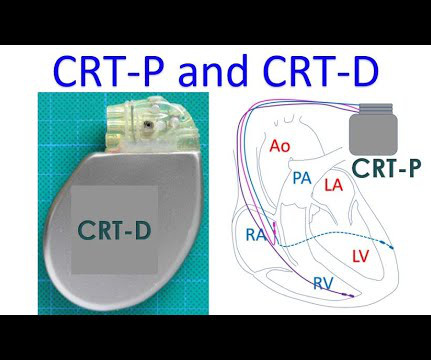
Moreover, the internal shocks given by CRT-D can be painful, though essential to save life in case of fast ventricular tachycardia or fibrillation. Naturally, the longevity of CRT-D battery is lesser than that of CRT-P. CRT-D is needed in some heart failure patients who have a high risk of life threatening ventricular arrhythmias.
One is ventricular tachycardia with regular retrograde activation. Especially, in patients with rheumatic fever, PR interval is prolonged and there is sinus tachycardia. Second is junctional tachycarida. Then sometimes, prolonged first degree AV block. Suppose this is a little more prolonge, this P will come here.
In that study commonest ECG abnormalites were QTc prolongation followed by brady/tachycardia and then ST segment deviations [3]. In a study involving patients with supratentorial hemorrhage, they found that ECG changes were more common in basal ganglia and thalamic bleeds rather than in lobar bleeds.
This is one important cause of supraventricular tachycardia in Ebstein’s anomaly. So, in fact, if an ECG of a person with Ebstein’s anomaly does not show incomplete right bundle branch block pattern, then an accessory pathway is suspected. Electrophysiological study will show that, and this pathway can be ablated.
ECG in a person with persistent anginal pain for the past several hours showing significant ST segment depression anterolateral leads along with sinus tachycardia. ST segment elevation is noted in aVR. Such a pattern is consistent with significant left main coronary artery stenosis.
Second reason is that they have more of ventricular fibrillation than ventricular tachycardia. Secondly, that is, in those who are having basic left ventricular dysfunction, after myocardial infarction, residual LV dysfunction.
We organize all of the trending information in your field so you don't have to. Join thousands of users and stay up to date on the latest articles your peers are reading.
You know about us, now we want to get to know you!
Let's personalize your content
Let's get even more personalized
We recognize your account from another site in our network, please click 'Send Email' below to continue with verifying your account and setting a password.


Let's personalize your content