This site uses cookies to improve your experience. To help us insure we adhere to various privacy regulations, please select your country/region of residence. If you do not select a country, we will assume you are from the United States. Select your Cookie Settings or view our Privacy Policy and Terms of Use.
Cookie Settings
Cookies and similar technologies are used on this website for proper function of the website, for tracking performance analytics and for marketing purposes. We and some of our third-party providers may use cookie data for various purposes. Please review the cookie settings below and choose your preference.
Used for the proper function of the website
Used for monitoring website traffic and interactions
Cookie Settings
Cookies and similar technologies are used on this website for proper function of the website, for tracking performance analytics and for marketing purposes. We and some of our third-party providers may use cookie data for various purposes. Please review the cookie settings below and choose your preference.
Strictly Necessary: Used for the proper function of the website
Performance/Analytics: Used for monitoring website traffic and interactions
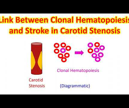
About a fifth of all ischemic strokes are attributed to embolization of ruptured atherosclerotic plaque from carotid arterial stenosis. But it has been difficult to predict which person with asymptomatic carotid artery stenosis is likely to progress to symptomatic carotid disease and stroke. J Am Coll Cardiol. doi: 10.1016/j.jacc.2024.03.389.
Such a pattern is consistent with significant left main coronary artery stenosis. Angiography done after initial stabilization showed severe stenosis of distal left main coronary artery. ST segment elevation is noted in aVR. Clinical evaluation and X-Ray chest showed features of pulmonary edema.
This is a simplified equation routinely used in Doppler echocardiography for pressure gradient across valves while quantifying severity of stenosis. Velocity proximal to the stenosis being much smaller, is ignored. In the simplified equation, only the distal velocity is taken into consideration.
Tetralogy of Fallot TOF with pulmonary atresia Pulmonary atresia with intact interventricular septum Tricuspid atresia Double outlet right ventricle Transposition of great arteries with ventricular septal defect and pulmonary stenosis Ebstein’s anomaly of tricuspid valve In DORV and tricuspid atresia, there are also variants with increased pulmonary (..)
Patients were stratified based on diabetes status, and various outcome measures were assessed using Kaplan-Meier event rates and Cox model hazard ratios.
D-Transposition of great arteries Double outlet right ventricle without pulmonary pulmonary stenosis Taussig-Bing anomaly Total anomalous pulmonary venous return Truncus arteriosus Single ventricle (double inlet ventricle, univentricular heart)
One is ventricular septal defect, second is overriding aorta, third is pulmonary stenosis, usually right ventricular outflow tract stenosis and associated right ventricular hypertrophy. Pulmonary stenosis, which is usually right ventricular outflow tract stenosis. As the name implies, there are four defects.
Coronary angiography gives a visual impression about the severity of the stenosis. But it need not imply the actual functional significance of the stenosis in terms of flow physiology. A downside of the study was that it had included lesions of 50 to 79% stenosis also. identified physiologically significant stenosis.
A study published in Radiology , the flagship journal of the Radiological Society of North America evaluated both in vivo and in vitro coronary stenosis using ultrahigh-spatial-resolution photon counting detector CT reconstructions [1]. Reconstructions were at standard resolution of 0.6 Section increments were 0.4 mm and 0.1
SCAPE is an acronym for sympathetic crash acute pulmonary edema, which can typcially occur in Pickering syndrome with renal artery stenosis [1]. Another term for transient acute pulmonary edema which occurs in renal artery stenosis is flash pulmonary edema.
Graft material has the disadvantage that it will not grow as the baby grows and can lead to supravalvar pulmonary stenosis later, one of the delayed complications of arterial switch. This is diagrammatic representation of stenosis of pulmonary artery at the site where it has been repaired.
Transcatheter aortic valve replacement (TAVR) is increasing in popularity for symptomatic severe aortic stenosis. No calcifications in the artery causing vascular stenosis. Contralateral or left atherosclerosis in the carotid artery with significant stenosis or occlusion was a contraindication for this approach.
In the geometry, the size of the plaque, its relationship to luminal stenosis, arterial remodeling and eccentricity can be evaluated. Transient ischemia may occur while negotiating tight stenosis or small vessels. Plaque morphology assessment with IVUS Plaque morphology can be assessed in terms of its geometry and echogenicity.
Colour Doppler echocardiography is very useful in giving a quick visual assessment of regurgitation and stenosis of heart valves. Stenosis of the valves can be made out by the reduced opening at the time when it is supposed to be open. All high velocity flows across the diseased valves will be shown as mosaic jets.
Bicuspid leaflets are likely to have both stenosis and regurgitation and both can progress as age advances, so that they may present with aortic stenosis most often in the older age group. And then, this is likely to be regurgitant also.
Just as water logging occurs in the catchment area of a dam after a heavy rain, fluid collects in the lungs if the valve between the left upper and lower chambers of the heart (mitral valve) is narrowed (mitral stenosis).
So that is why we see straightening of left border, typically heard of in mitral stenosis with left atrial enlargement and mild pulmonary hypertension. Normally, the main pulmonary artery segment will be concave and left atrial appendage region also will be not prominent.
Fetal aortic valvuloplasty is considered for fetuses with severe valvar aortic stenosis and echocardiographic features suggesting a risk of progression to hypoplastic left heart syndrome. Though surgical options are available for infants with hypoplastic left heart syndrome, morbidity and mortality are high.
SMART 4 ( NCT04722250 ) studied patients with severe aortic stenosis and a small aortic annulus who underwent transcatheter aortic valve replacement (TAVR). The study concluded that among low-risk patients with severe aortic stenosis, TAVI is as effective as SAVR in terms of the composite outcome of death or stroke at 1 year.
The Y descent is shallow in tricuspid stenosis, and absent in cardiac tamponade. Right atrial hypertrophy as in tricuspid stenosis, pulmonary stenosis and pulmonary hypertension. X descent, X prime descent and Y descent. The X prime descent is absent in tricuspid regurgitation in which, C and V waves fuse together.
When there is associated mitral stenosis, the colour Doppler jet of mitral flow merges with that of aortic regurgitation in the left ventricle as both occur in diastole. m/sec, indicating the absence of any associated aortic stenosis. This is because the regurgitant flow into the left ventricle is towards the transducer in this view.
Relative contraindications for HUTT include: Severe left ventricular outflow obstruction Critical mitral stenosis Severe proximal coronary artery disease Severe cerebrovascular disease
Potentialy serious, though rare complications of ablation for atrial fibrillation are phrenic nerve injury resulting in diaphragmatic paralysis, pulmonary vein stenosis and esophagial injury resulting in catastrophic atrio-esophageal fistula.
Here is an example of a continuous wave Doppler interrogation of aortic stenosis and regurgitation. Most of the abnormal jets across abnormal valves and those across a ventricular septal defect and patent ductus arteriosus need either continuous wave or high pulse repetition Doppler for analysis due to the high velocity.
So it will not produce a true LV to aorta pullback tracing, which is required in cases like aortic stenosis. When the tip is in the left ventricle, this region will be in the aorta sometimes. For that you will have to use a catheter without side holes like this, like a multi-purpose catheter or some other catheter you have to use.
Planimetry of mitral valve area can be obtained in parasternal short axis view in case of mitral stenosis. Right ventricular cavity is elliptical in this view and left ventricular cavity is circular. Wall motion of the left ventricle can be assessed in this view also.
If a nominal right atrial pressure of 10 mm Hg is added to it, right ventricular pressure and indirectly the pulmonary artery systolic pressure are obtained, in the absence of pulmonary stenosis. If there is high right atrial pressure with elevated jugular venous pressure, 15 or 20 mm Hg may have to be added instead of 10 mm Hg.
We organize all of the trending information in your field so you don't have to. Join thousands of users and stay up to date on the latest articles your peers are reading.
You know about us, now we want to get to know you!
Let's personalize your content
Let's get even more personalized
We recognize your account from another site in our network, please click 'Send Email' below to continue with verifying your account and setting a password.


Let's personalize your content