This site uses cookies to improve your experience. To help us insure we adhere to various privacy regulations, please select your country/region of residence. If you do not select a country, we will assume you are from the United States. Select your Cookie Settings or view our Privacy Policy and Terms of Use.
Cookie Settings
Cookies and similar technologies are used on this website for proper function of the website, for tracking performance analytics and for marketing purposes. We and some of our third-party providers may use cookie data for various purposes. Please review the cookie settings below and choose your preference.
Used for the proper function of the website
Used for monitoring website traffic and interactions
Cookie Settings
Cookies and similar technologies are used on this website for proper function of the website, for tracking performance analytics and for marketing purposes. We and some of our third-party providers may use cookie data for various purposes. Please review the cookie settings below and choose your preference.
Strictly Necessary: Used for the proper function of the website
Performance/Analytics: Used for monitoring website traffic and interactions
As the medical community delves deeper into the intricacies of ANOCA, the study provides a compelling narrative for clinicians and researchers alike, aiming to decipher the complexities of myocardial bridges and their role in exercise-induced ischemia. Original article: Sinha A et al. Circ Cardiovasc Interv.
The images demonstrate a moderately extensive, mildly severe reversible defect in the mid and distal anterior/anterolateral wall consistent with ischemia. Which of the vessels is likely the culprit vessel causing the ischemia? The patient also had a cardiac CT for calcium scoring pictured below. The technologist performs a MUGA scan.
Results revealed ischemia in one cardiac territory in 80% of patients, two territories in 17%, and three territories in 2%. The investigators concluded that in patients with stable angina and minimal medication, confirmed ischemia, and randomized to PCI, the procedure led to better angina outcomes compared to the placebo.
Monomorphic ventricular tachycardia in the setting of acute myocardial ischemia can also be treated by intravenous lignocaine bolus followed by infusion. Predisposing causes for ventricular tachycardia like ischemia and electrolyte imbalance has to be treated simultaneously to prevent recurrence.
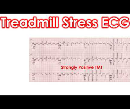
The recording in early phase of recovery at 1 minute, shows very little ST segment depression, making us suspect further whether the earlier recording was really due to myocardial ischemia. But the ST segment is down sloping in inferior leads.
indicates inducible ischemia while an FFR above 0.80 excludes ischemia in 90% of cases. There is a strong correlation between FFR and inducible myocardial ischemia. It recalculates SYNTAX score by incorporating ischemia producing lesions determined by FFR. Normal FFR is 1.0 and an FFR below 0.75 An FFR below 0.75
Mechanism is thought to be due to sustained sympathetic stimulation, probably caused by dysfunction of insular cortex resulting in reversible neurogenic damage to the myocardium which could include contraction bands and subendocardial ischemia [2]. Serial measurements of cardiac enzymes were normal in that case.
Transient ischemia may occur while negotiating tight stenosis or small vessels. But these major complications occur usually during interventions rather than diagnostic IVUS evaluations. Vasospasms are usually transient and respond to intracoronary nitroglycerine.
Pediatric exercise testing may be used for evaluation of various disorders of cardiac rhythm rather than for inducible ischemia as in adults. Discussion on pediatric exercise testing. In a child with suspected sinus node dysfunction, chronotropic incompetence from sinus node dysfunction can be assessed by exercise testing.
That is, there is renal ischemia due to clogging of capillaries, which leads to increased erythropoietin secretion and this causes decompensated erythrocytosis. And it was described from the very first report of Eisenmenger syndrome. That has already been discussed. Decompensated erythrocytosis is something which we see very often.
Absence of brachiocephalic artery occlusion which will increase the risk of intraprocedural cerebral ischemia 3. Brachiocephalic artery diameter 8 mm or more on CT so that 18-F sheath could be inserted and blood flow could occur through the gap between the sheath and the vessel 2. No calcifications in the artery causing vascular stenosis.
ST segment elevation in aVR in proximal LAD occlusion before first septal is thought to be due to transmural ischemia of the basal part of the septum. ST elevation in aVR greater than or equal to that in V1 distinguished left main stenosis from left anterior descending coronary artery stenosis with 81% sensitivity and 80% specificity.
Automatic ventricular tachycardia can occur in acute ischemia, electrolyte imbalance and with increased sympathetic tone. Ventricular tachycardia can be classified according to the mechanism of genesis into reentrant, automatic and triggered activity. Triggered activity can be early and delayed afterdepolarization.
They include myocardial ischemia, acute pericarditis, pulmonary embolism, external compression due to mass over the right ventricular outflow tract region, and metabolic disorders like hyper or hypokalemia and hypercalcemia. These are the conditions which have to be considered or excluded as they can sometimes manifest Brugada pattern on ECG.
The highly impactful International Study of Comparative Health Effectiveness With Medical and Invasive Approaches ( ISCHEMIA ) trial investigated the effectiveness of invasive (INV) versus conservative (CON) strategies for managing stable coronary artery disease. EuroIntervention. 2024 Mar 5:EIJ-D-24-00011.
We organize all of the trending information in your field so you don't have to. Join thousands of users and stay up to date on the latest articles your peers are reading.
You know about us, now we want to get to know you!
Let's personalize your content
Let's get even more personalized
We recognize your account from another site in our network, please click 'Send Email' below to continue with verifying your account and setting a password.


Let's personalize your content