This site uses cookies to improve your experience. To help us insure we adhere to various privacy regulations, please select your country/region of residence. If you do not select a country, we will assume you are from the United States. Select your Cookie Settings or view our Privacy Policy and Terms of Use.
Cookie Settings
Cookies and similar technologies are used on this website for proper function of the website, for tracking performance analytics and for marketing purposes. We and some of our third-party providers may use cookie data for various purposes. Please review the cookie settings below and choose your preference.
Used for the proper function of the website
Used for monitoring website traffic and interactions
Cookie Settings
Cookies and similar technologies are used on this website for proper function of the website, for tracking performance analytics and for marketing purposes. We and some of our third-party providers may use cookie data for various purposes. Please review the cookie settings below and choose your preference.
Strictly Necessary: Used for the proper function of the website
Performance/Analytics: Used for monitoring website traffic and interactions
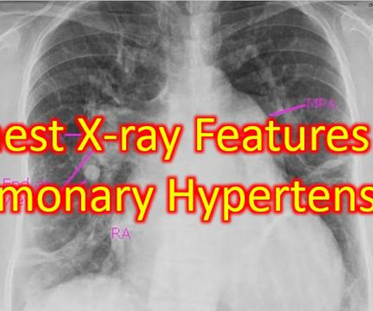
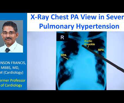
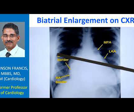
Couple of chest X-rays showing serial changes in pulmonary hypertension over the years and another with features of severe pulmonary hypertension. The post Chest X-ray Features of Pulmonary Hypertension appeared first on All About Cardiovascular System and Disorders.
Transcript of the video: Now we will discuss a chest X-ray showing antler sign in pulmonary venous hypertension. So, the shape of the upper lobe vessels in venous hypertension, pulmonary venous hypertension, will be resembling the antlers of the stag. For a change, first we will see the picture of a stag, with antlers.
It can occur in severe pulmonary hypertension, there can be right atrial enlargement, as the severe hypertrophy of the right ventricle causes elevation of right ventricular end diastolic pressure, and later on right atrial enlargement. In VSD Eisenmenger and PDA Eisenmenger, you would not expect right atrial enlargement.
This multicenter open-label, blinded-outcome, randomized trial ( NCT04030234 ), offers new insights into hypertension management. No significant differences were observed in hypotension, electrolyte abnormality, injurious falls, or acute kidney injury between the groups.
Nuclear Cardiology See if you’re ready for the Nuclear Cardiology boards by answering these hard-hitting sample questions plucked from our very own nuclear cardiology question bank. A 48 year-old female with hypertension, hyperlipidemia, chronic low back pain, and bilateral lower extremity neuropathy.
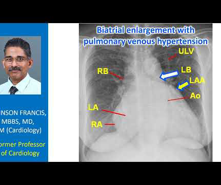
So that is why we see straightening of left border, typically heard of in mitral stenosis with left atrial enlargement and mild pulmonary hypertension. When there is gross pulmonary hypertension, instead of these being straight over here, it will form a bulge over here.
Transcript of the video: Eisenmenger syndrome is an important complication of large left to right shunts which develop later due to development of pulmonary vascular obstructive disease and severe pulmonary hypertension. Some even say that those with Eisenmenger in ASD are really those who are destined for primary pulmonary hypertension.
Among the 285 former players in the study, of which over two thirds were African American, prevalence of hypertension was 89.8%, though only 37.5% Hypertension was a significant predictor of clinically relevant structural abnormalities on transthoracic echocardiogram. were aware of it.
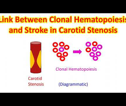
It has been mentioned that risk due to clonal hematopoiesis is equal in magnitude to conventional risk factors like smoking, hypertension, dyslipidemia and diabetes. Those having these mutations have increased vascular inflammation before it progresses to malignancies and double the risk of ischemic stroke.
One, the flow may be increased in the long run, leading to pulmonary hypertension and pulmonary vascular disease. So this reduces the chance of future development of pulmonary hypertension and also reduces the impairment of the arm blood supply. This will improve the pulmonary blood flow and relieve cyanosis.
In fact, the technical name for high blood pressure is hypertension! One of the criteria for a good blood pressure recording is that you should not have taken caffeinated drinks within half an hour of the measurement. Reducing stress is another method of reducing blood pressure.
But then that is not a stroke but called as hypertensive encephalopathy (brain disease due to high blood pressure). Even without a bleed, brain function can be altered due to high blood pressure, causing alteration in the level of consciousness.
The cyanosis in Ebstein’s anomaly, is usually not due to pulmonary hypertension, but because tricuspid regurgitation jet is directed across the atrial septal defect. Ebstein’s anomaly may be associated with atrial septal defect or a patent foramen ovale, in about 50% of cases.
Guidelines on hypertension (high blood pressure) generally recommend measurement of blood pressure in both arms in the initial visit. They also suggest that the arm with higher blood pressure recording should be used to record blood pressure in subsequent visits.
Right atrial hypertrophy as in tricuspid stenosis, pulmonary stenosis and pulmonary hypertension. But in a VSD with pulmonary hypertension A wave is not prominent. We noted that prominent V waves or CV waves can occur in tricupsid regurgitation. A prominent A wave can occur when the atrium is hypertrophied.
in hypertensives are some of the features. Hypertensive heart disease is an important differential diagnosis, but SAM is rare in this situation and there is evidence of greater diastolic dysfunction in HCM. Septal thickness is often 4-6 mm more than normal. in normotensives and more than 1.5
Tricuspid regurgitation jet velocity and pulmonary regurgitation end diastolic velocity indicating pulmonary hypertension are also taken as surrogates of left atrial pressure in the absence of pulmonary disease. Doppler interrogation of mitral valve is usually done from the apex through the apical four chamber view.
Inclusion criteria were AF diagnosed within 12 months of enrollment, age above 75 years, previous transient ischemic attack or stroke or those who had two of the following criteria: age above 65 years, female gender, heart failure, hypertension, diabetes mellitus, severe coronary artery disease, chronic kidney disease stage 3 or 4, left ventricular (..)
Adiposity activates inflammatory cytokines, enhances risk of hypertension, dyslipidemia and diabetes mellitus. Less quantities of healthier food may be consumed in the diet by those with higher intake of sugar sweetened beverages. Inflammatory cytokines causing unstable plaques lead on to cardiovascular events [1].
Most common method of assessment of pulmonary hypertension by Doppler echocardiography is by using: A: Forward velocity across the tricuspid valve B: Reverse velocity across the tricuspid valve C: Forward velocity across the pulmonary valve D: Reverse velocity across the mitral valve Correct answer: B: Reverse velocity across the tricuspid valve Reverse (..)
We organize all of the trending information in your field so you don't have to. Join thousands of users and stay up to date on the latest articles your peers are reading.
You know about us, now we want to get to know you!
Let's personalize your content
Let's get even more personalized
We recognize your account from another site in our network, please click 'Send Email' below to continue with verifying your account and setting a password.


Let's personalize your content