This site uses cookies to improve your experience. To help us insure we adhere to various privacy regulations, please select your country/region of residence. If you do not select a country, we will assume you are from the United States. Select your Cookie Settings or view our Privacy Policy and Terms of Use.
Cookie Settings
Cookies and similar technologies are used on this website for proper function of the website, for tracking performance analytics and for marketing purposes. We and some of our third-party providers may use cookie data for various purposes. Please review the cookie settings below and choose your preference.
Used for the proper function of the website
Used for monitoring website traffic and interactions
Cookie Settings
Cookies and similar technologies are used on this website for proper function of the website, for tracking performance analytics and for marketing purposes. We and some of our third-party providers may use cookie data for various purposes. Please review the cookie settings below and choose your preference.
Strictly Necessary: Used for the proper function of the website
Performance/Analytics: Used for monitoring website traffic and interactions
When thyroid function is increased, heart rate increases and the work load of the heart increases. In severe cases heartfailure may occur. While in low output heartfailure the extremities are cold, in high output failure due to increased thyroid function, the extremities of the limbs are warm.
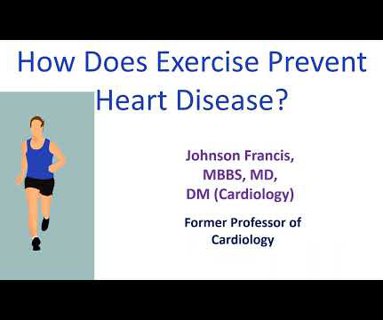
Plaque regression can be demonstrated by ultrasound evaluation of the carotids which are easily accessible. Lower blood pressures mean lower workload for the heart and lower risk of left ventricular hypertrophy and heartfailure. Regular exercise can bring down the blood pressure in the long run.
One situation is decompensated advanced systolic heartfailure with large left ventricle. J Cardiovasc Ultrasound. J Cardiovasc Ultrasound. Measurement of E’ is useful in differentiation of pseudo normalization in the mitral inflow from the normal pattern. Ha J et al. 2009 Sep;17(3):86-95. Park JH et al. Møller JE et al.
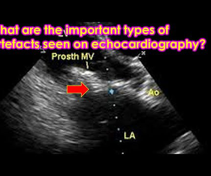
Lower energy side beams of ultrasound known as sidelobes can reflect off lateral structures and get mapped to the central image, causing sidelobe artefacts. Multiple reflections from strong reflectors of ultrasound like pericardium and pleura can cause mirror images of structures being imaged.
We organize all of the trending information in your field so you don't have to. Join thousands of users and stay up to date on the latest articles your peers are reading.
You know about us, now we want to get to know you!
Let's personalize your content
Let's get even more personalized
We recognize your account from another site in our network, please click 'Send Email' below to continue with verifying your account and setting a password.


Let's personalize your content