This site uses cookies to improve your experience. To help us insure we adhere to various privacy regulations, please select your country/region of residence. If you do not select a country, we will assume you are from the United States. Select your Cookie Settings or view our Privacy Policy and Terms of Use.
Cookie Settings
Cookies and similar technologies are used on this website for proper function of the website, for tracking performance analytics and for marketing purposes. We and some of our third-party providers may use cookie data for various purposes. Please review the cookie settings below and choose your preference.
Used for the proper function of the website
Used for monitoring website traffic and interactions
Cookie Settings
Cookies and similar technologies are used on this website for proper function of the website, for tracking performance analytics and for marketing purposes. We and some of our third-party providers may use cookie data for various purposes. Please review the cookie settings below and choose your preference.
Strictly Necessary: Used for the proper function of the website
Performance/Analytics: Used for monitoring website traffic and interactions
There is a regular narrow complex tachycardia. Thus, it is supraventricular tachycardia. It is important to remember that SVT includes Sinus Tachycardia! Sometimes even Wide Complex Tachycardia is Sinus. See this case in which Lewis leads were necessary to figure this out: Wide Complex Tachycardia.
I find AV dissociation in VT to be very difficult to differentiate from artifact, as there are always random blips on tachycardia tracings. Read this post: Idiopathic Ventricular Tachycardias for the EM Physician 2. The 15th beat (2nd beat of V1-V3) appears to be a fusion beat , which is all but diagnostic of VT.
Discussion on pediatric exercise testing. Pediatric exercise testing may be used for evaluation of various disorders of cardiac rhythm rather than for inducible ischemia as in adults. In a child with suspected sinus node dysfunction, chronotropic incompetence from sinus node dysfunction can be assessed by exercise testing.
"CV Sports Chat" is an interview series including expert discussions relative to sports and exercise cardiology and the health care management of athletes.
She was awake, alert, well perfused, with normal mental status and overall unremarkable physical exam except for a regular tachycardia, possible rales at both bases, some mild RUQ abdominal tenderness. Thus, I believe it is a regular, monomorphic, wide complex tachycardia. Or it could simply still be classic VT. RVEF 100 ml/m2.
Catecholaminergic polymorphic ventricular tachycardia (CPVT) is a rare disorder presenting as exercise-induced ventricular arrhythmias, rarely associated with mutations in triadin (TRDN).
This strip was obtained: Apparent Wide Complex Tachycardia at a rate of 280 What do you think? Because the patient was exercising, which increases sympathetic tone, facilitating AV conduction. Troponins 34>33>43, likely secondary to myocardial injury from tachycardia. What do you want to do? Why such rapid AV conduction?
By identifying patterns, users can understand how their heart responds to exercise, stress, or relaxation. Tracking Physical Activity and Exercise Physical activity is vital for maintaining heart health, and wearable tech provides detailed metrics on steps taken, calories burned, and active minutes.
The primary end point was syncope recurrence, and the secondary end point was the reduction of the ventricular arrhythmia score during exercise testing. Twenty‐six PVT/ventricular fibrillation–triggering PVCs were identified for ablation. Induction of nontriggering PVCs after ablation is associated with a higher risk of syncope recurrence.
Here is her ED ECG: Here is the ED physician's interpretation: IMPRESSION UNCERTAIN REGULAR RHYTHM, wide complex tachycardia, likely p-waves. LEFT BUNDLE BRANCH BLOCK [120+ ms QRS DURATION, 80+ ms Q/S IN V1/V2, 85+ ms R IN I/aVL/V5/V6] Comparison Summary: LBBB and tachycardia are new. This is clearly ventricular tachycardia.
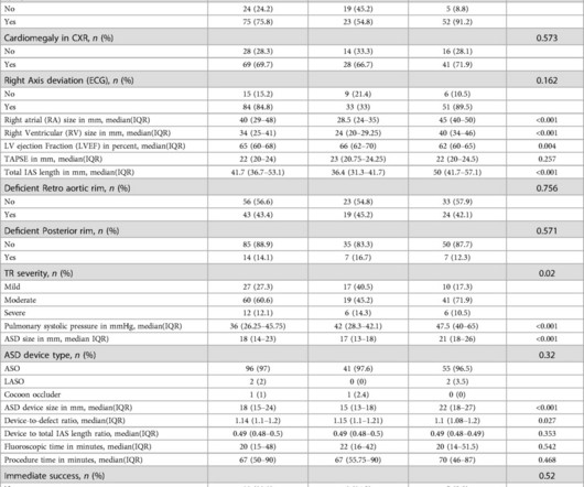
When it is large and hemodynamically significant, it can cause symptoms such as dyspnea, exercise intolerance, and palpitations. and paroxysmal supraventricular tachycardia (SVT) (5.3%), respectively. IntroductionSecundum Atrial septal defect (ASD) is the most common type of ASD. and Group 2: 2.5 (IQR IQR 22.5); P-value=0.111].
Initial ECG in the ED: Presenting ECG : Wide-complex tachycardia at a rate about 200. This is overwhelmingly likely to be ventricular tachycardia, even if only age and medical history are considered. Nevertheless, the widths of both the QRS complex and the RS duration are similar in both the old ECG and the tachycardia.
Left ventricular summit VT is a type of benign idiopathic adenosine-sensitive VT that is caused by triggered activity and can be precipitated by excess catecholamines, such as during exercise. Unlike other outflow tract VTs, LV summit VT can be challenging to ablate because of its intramural or epicardial origin.
Sinus tachycardia – sinus rhythm above 100 bpm is a sinus tachycardia. In healthy individuals occurs during exercising or strong emotions. Ventricular tachycardia – more than 7 consecutive complexes originating from ventricles at a rate of > 100 bpm. Usually does not exceed 160 bpm.
Abstract Introduction We report the case of a 37-year-old male athlete, who developed during exercise atrial and ventricular arrhythmias. Conclusions The findings in our patient may suggest some increased ventricular excitability using programmed ventricular stimulation in CASQ2 polymorphic ventricular tachycardia patients.
With increased genetic testing, sudden cardiac arrest (SCA) survivors who carry variants in the cardiac ryanodine receptor (RYR2) gene but do not have the pathognomonic findings of catecholaminergic polymorphic ventricular tachycardia (CPVT) on exercise testing were identified.
Exercise test would also have been reasonable. His symptoms of chest pain and shortness of breath were attributed to an asthma exacerbation during exercise. Read about "exercise induced cardiac troponin elevations" here. Potential Adverse Cardiovascular Effects From Excessive Endurance ExerciseExercise Is Medicine?
Here is his 12-lead: There is a wide complex tachycardia with a rate of 257, with RBBB and LPFB (right axis deviation) morphology. Read about Fascicular VT here: Idiopathic Ventricular Tachycardias for the EM Physician Case Continued He was completely stable, so adenosine was administered. See Learning point 1 below. Arch Intern Med.
The main secondary study endpoints are all-cause mortality, cardiovascular mortality, incidence of implantable cardioverter-defibrillator (ICD) therapy, hospitalizations, quality of life, time to first ICD therapy, number of device-detected ventricular tachycardia/ventricular fibrillation episodes, left ventricular function, and exercise tolerance.
This usually represents posterior OMI, but in tachycardia and especially after cardiac arrest, this could simply be demand ischemia, residual subendocardial ischemia due to the low flow state of the cardiac arrest. This prompted cath lab activation. On arrival to the ED, this ECG was recorded: What do you think?
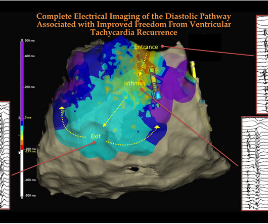
The learning point is that, in VT ablation, looking for anatomical diastolic tracts and its electrical activity becomes a key exercise. Luca Rosario Limite , et al Complete Electroanatomic Imaging of the Diastolic Pathway Is Associated With Improved Freedom From Ventricular Tachycardia Recurrence Circ Arrhythm Electrophysiol.
She underwent exercise echocardiogram in mid October where she exercised for nearly 7 minutes on the standard Bruce protocol and had typical anginal pain and shortness of breath. She has been experiencing progressively worsening exertional dyspnea and chest tightness mostly when climbing up flights of stairs since early September.
Typically — these arrhythmias are induced by exercise or intense emotional states ( ie, associated with increased catecholamine discharge ). As a result — ETT ( Exercise Treadmill Testing ) can be used to elicit BDVT, and therefore help to diagnosie CPVT. As a result — the resting ECG tends to be normal.
This is one important cause of supraventricular tachycardia in Ebstein’s anomaly. Ebstein’s anomaly may present with a murmur for evaluation in the pediatric age group or in adults with arrhythmias or heart failure with cyanosis and exercise intolerance.
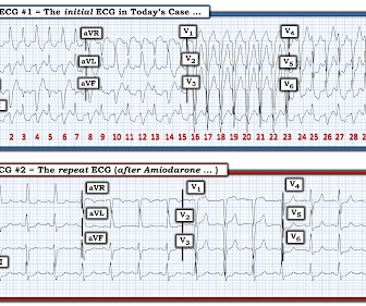
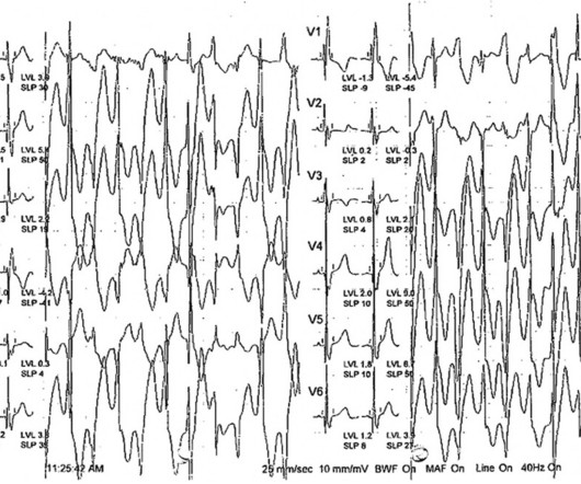
The ECG in Figure-1 was obtained from a previously healthy middle-aged man — who while performing his regular exercise routine, developed "slight" chest discomfort and "palpitations". Given the rapid rate of the tachycardia and the amorphous shape of the QRS — the decision was made to sedate the patient and cardiovert.
Other parameters which are thought to convey risk in Brugada syndrome are Tpeak-Tend >100 ms in chest leads, early repolarization pattern in inferior leads, post exercise ST segment elevation and diurnal burden of Type 1 ECG pattern on Holter monitoring [4]. Opinion is divided on the need for electrophysiology study. mV or R/q ≥ 0.75.
BackgroundCatecholaminergic polymorphic ventricular tachycardia (CPVT) is a rare inherited arrhythmia disorder characterized by ventricular arrhythmia triggered by adrenergic stimulation.Case presentationA 9-year-old boy presented with convulsions following physical exertion. Genetic testing revealed a pathogenic variant of RYR2:c.720G>A
Introduction:Postural orthostatic tachycardia syndrome (POTS) is a chronic syndrome with symptoms of orthostatic intolerance, tachycardia, and exercise intolerance. At peak exercise, there was no difference in respiratory exchange ratio (1.18±.2 Resting HR was lower in male patients (76±17 vs 85±18 bpm, p<0.05).
The abnormal heart rhythms can further lead to death because of ventricular tachycardia and ventricular fibrillation. Get some exercise regularly. Exercise offers numerous advantages, including strengthening the heart and improving circulation. Besides, excessive stress can also act as a “trigger” for a heart attack.
Previously healthy, taking no medication and exercising regularly. No anginal symptoms asymptomatic during physical exercise. During observation in the ED the patient had multiple self-terminating runs of Non-Sustained monomorphic Ventricular Tachycardia (NSVT). Below in Figure-1 is this patient's admission ECG.
We organize all of the trending information in your field so you don't have to. Join thousands of users and stay up to date on the latest articles your peers are reading.
You know about us, now we want to get to know you!
Let's personalize your content
Let's get even more personalized
We recognize your account from another site in our network, please click 'Send Email' below to continue with verifying your account and setting a password.


Let's personalize your content