This site uses cookies to improve your experience. To help us insure we adhere to various privacy regulations, please select your country/region of residence. If you do not select a country, we will assume you are from the United States. Select your Cookie Settings or view our Privacy Policy and Terms of Use.
Cookie Settings
Cookies and similar technologies are used on this website for proper function of the website, for tracking performance analytics and for marketing purposes. We and some of our third-party providers may use cookie data for various purposes. Please review the cookie settings below and choose your preference.
Used for the proper function of the website
Used for monitoring website traffic and interactions
Cookie Settings
Cookies and similar technologies are used on this website for proper function of the website, for tracking performance analytics and for marketing purposes. We and some of our third-party providers may use cookie data for various purposes. Please review the cookie settings below and choose your preference.
Strictly Necessary: Used for the proper function of the website
Performance/Analytics: Used for monitoring website traffic and interactions
Aims Exercise testing remains underused in patients with aortic stenosis (AS), partly due to concerns about an exercise-induced drop in systolic blood pressure (SBP). We aimed to study the SBP response to exercise in patients with severe symptomatic AS prior to surgery and 1 year postoperatively.
Valvular heart disease, including calcific or degenerative aortic stenosis (AS), is increasingly prevalent among the older adult population. Exercise during the preoperative waiting period may be safe and effective in most patients with severe AS.
BackgroundThe potential impact of exercise on valvular function and aortic diameters in patients with a bicuspid aortic valve remains unclear. Echocardiography was used to assess aortic stenosis or aortic regurgitation and to measure diameters at the sinuses of Valsalva and ascending aorta. Aortic dilatation was defined as aZ‐score ≥2.
Since the pathologist does not know the original cross-sectional area of the artery or the amount of compensatory enlargement of the artery from evaluation of a single cross section of the artery at a site of stenosis, the degree of luminal narrowing of that segment cannot be determined. These are typical findings at sites of plaque rupture.
Reducing the high risk of recurrent stroke in patients with symptomatic intracranial atherosclerotic stenosis (sICAS) has proven to be challenging, but aggressive medical management, with intensive risk factor control and antithrombotic therapy, has been shown to be beneficial. Stroke, Volume 55, Issue 2 , Page 335-343, February 1, 2024.
Optimal administration of exercise or intravenous drugs may reveal hemodynamic abnormalities under stress without posing an invasive risk. Therefore, the implementation of stress echocardiography is recommended for determining interventional indications and risk stratification in mitral regurgitation and aortic stenosis.
More than 90% of the Lp(a) level is influenced by variations in the genes controlling the Lp(a) particle production, 2 in which lifestyle interventions such as diet and exercise have no significant impact. 2022 Aug, 80 (9) 934946 Kronenberg F.
Transcatheter aortic valve replacement (TAVR) is a relatively new treatment method for aortic stenosis (AS) and has been demonstrated to be suitable for patients with varying risk levels.
The emergency department of Liaocheng People's Hospital in Shandong Province admitted one patient with OHCA in August 2021, who suddenly suffered a loss of consciousness and cardiac arrest during exercise after dinner. Fortunately, there was no obvious stenosis in the right coronary artery.
Sent by anonymous A man in his 40s with no previous heart disease presented within 30 minutes of onset of acute chest pain that started while exercising. Angiogram findings included: 95% mid RCA stenosis with occluded distal right PDA secondary to thrombus (peristent OMI). Written by Pendell Meyers with edits by Smith.
On this visit, he expressed worsening exercise tolerance, new orthopnea, and he told his provider that the omeprazole did not relieve any symptoms. The red arrow shows a roughly 80% stenosis of the proximal LAD. The blue arrow shows another stenosis of the LAD distal to the first diagonal branch of about 99%.
If you’ve been diagnosed with aortic stenosis, you might have come across the term TAVR. Understanding Aortic Stenosis The aortic valve regulates blood flow from your heart’s main pumping chamber to the rest of your body. In aortic stenosis, the valve leaflets stiffen and narrow, restricting blood flow.
She underwent exercise echocardiogram in mid October where she exercised for nearly 7 minutes on the standard Bruce protocol and had typical anginal pain and shortness of breath. Lesion on Dist RCA: 90% stenosis reduced to 0%. Baseline echocardiogram showed moderate LV systolic dysfunction with no wall motion abnormalities.
On follow up angiography, there was a large OM1 and small AV groove Cx/LPL visible as the vessel re-canalized LAD is noted to have diffuse 50% stenosis in the proximal segment and is occluded immediately beyond a small D1 RCA is a medium-large caliber vessel and supplies a medium rPDA, medium rPLA1, and three small rPLA branches. TIMI-0 flow.
Hiding behind the technicalities PCI demands reduction in percentage stenosis , resulting in pre-defined minimal luminal area (MLA), maximizing net luminal gain, & restoration of TIMI 3 flow in all three coronary arteries.These are the popular scientific parameters. Regular exercise equivalent to PCI (ESC 2009).Will
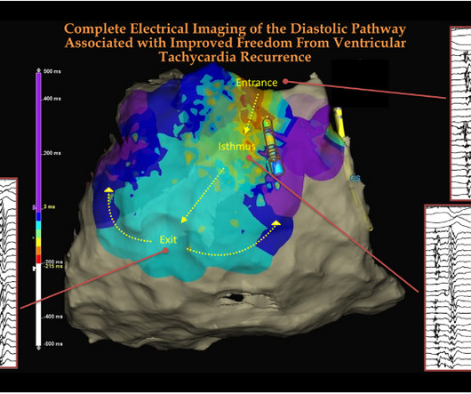
The learning point is that, in VT ablation, looking for anatomical diastolic tracts and its electrical activity becomes a key exercise. We can take a cue from the vintage clinical auscultation classes, where we ask the medical students to look for MDM (mid-diastolic murmur) in mitral stenosis in the left lateral posture in expiratory phase.
24: Joint American College of Cardiology/Journal of the American College of Cardiology Late-Breaking Clinical Trials (Session 402) Saturday, April 6 9:30 – 10:30 a.m.
It is prudent to understand, that even in systolic LV failure; it is the raised LVEDP that causes the symptoms and marks the limits of exercise capacity. We know a small ASD decompresses mitral stenosis, and the combination of ASD and MS, Lutembacher, is a well-known syndrome called Lutembacher.
He first noticed it while exercising. The LAD has diffuse disease with a few areas of moderate stenosis but no flow-limiting lesions. Over the next three days, he continued to have intermittent symptoms and therefore re-presented to the emergency room. The following ECG was obtained around midnight.
Furthermore, she denies any hydration since conclusion of exercise. There is ventricular hypertrophy in the absence of abnormal loading conditions, such as aortic stenosis, or hypertension, for example – of which the most common variant is Asymmetric Septal Hypertrophy.
As an exercise, lets calculate the equation for differentiating the ST elevation between benign early repolarization and LAD occlusion. With no other information other than the first ECG above, I texted this to Dr. Smith and he responded: ST elevation in lead V2 and terminal QRS distortion in V3. LAD occlusion. Great case.
BackgroundThe prognostic value of serial exercise echocardiography (EEC) in asymptomatic severe aortic stenosis is unknown. Journal of the American Heart Association, Ahead of Print.
Venn diagram highlighting the main similarities and differences between heart failure with preserved ejection fraction (HFpEF) and aortic stenosis with preserved ejection fraction (ASpEF). This study aimed to provide a non-invasive, comparative analysis of ASpEF versus HFpEF at rest and during exercise. vs. controls).
Objectives To develop a tool including exercise electrocardiography (ExECG) for patient-specific clinical likelihood estimation of patients with suspected obstructive coronary artery disease (CAD). In the CAD validation cohort, obstructive CAD was defined as >50% diameter stenosis on invasive coronary angiography.
The bilateral carotid artery stenosis (BCAS)-induced chronic cerebral hypoperfusion activates the NLRP inflammasome in brain tissue. This may be an important mechanism of action of physical exercise and C-RIC in VCID. Background and Purpose:Inflammation plays a significant role in acute and chronic cerebral ischemia.
One is ventricular septal defect, second is overriding aorta, third is pulmonary stenosis, usually right ventricular outflow tract stenosis and associated right ventricular hypertrophy. Pulmonary stenosis, which is usually right ventricular outflow tract stenosis. As the name implies, there are four defects.
The reason they have chosen to wear gym clothes is that they expect to do an exercise stress test as part of their assessment. Because if you are ‘getting your heart checked’ , you must do an exercise stress test, right? Do exercise stress tests tell you whether or not you have plaque in your coronary arteries?
This can lead to complications such as blocked, reduced, or backward blood flow through the heart chambers, causing shortness of breath, chest pain, fainting, and difficulty exercising. In more severe cases, the disease can lead to an aortic dissection, or tear in the aorta, a life-threatening condition.
Objective To characterise cardiac remodelling, exercise capacity and fibroinflammatory biomarkers in patients with aortic stenosis (AS) with and without diabetes, and assess the impact of diabetes on outcomes. to 4.00; p=0.037).
BACKGROUND:Recent guidelines redefined exercise pulmonary hypertension as a mean pulmonary artery pressure/cardiac output (mPAP/CO) slope >3 mm Hg·L−1·min−1. cm2underwent cardiopulmonary exercise testing with echocardiography. P=0.002), and age-, sex-, and height-based predicted peak exercise oxygen uptake (OR per SD, 0.59;P=0.007)
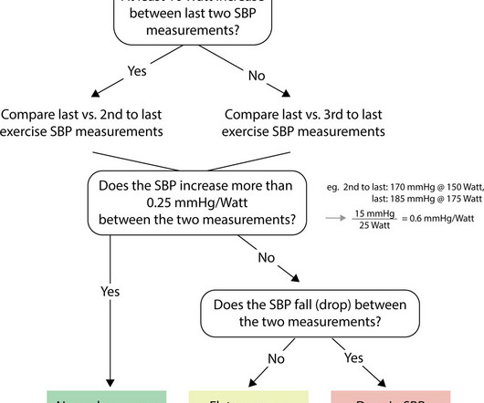
Background Knowledge about how patients with symptomatic aortic stenosis (AS) perform on cardiopulmonary exercise testing (CPET) is sparse. Since exercise testing in patients with symptomatic AS is not advised, submaximal parameters could be of special interest.
Below are some of the most common causes of heart murmurs: Increased Blood Flow: Innocent heart murmurs often occur when there is an increase in blood flow, such as during pregnancy, exercise, fever, or growth spurts in children. In adults, innocent murmurs can develop in response to temporary factors like pregnancy, fever, or exercise.
The therapeutics of coronary stenosis has become a technogical wonder, interwoven with statistical wordplay in the last few decades. Like, viability, scars, futility, and benefits of revascularization, imaging-assisted PCI, impact of PCI on exercise capacity, importance of risk factor management, etc.
We organize all of the trending information in your field so you don't have to. Join thousands of users and stay up to date on the latest articles your peers are reading.
You know about us, now we want to get to know you!
Let's personalize your content
Let's get even more personalized
We recognize your account from another site in our network, please click 'Send Email' below to continue with verifying your account and setting a password.


Let's personalize your content