This site uses cookies to improve your experience. To help us insure we adhere to various privacy regulations, please select your country/region of residence. If you do not select a country, we will assume you are from the United States. Select your Cookie Settings or view our Privacy Policy and Terms of Use.
Cookie Settings
Cookies and similar technologies are used on this website for proper function of the website, for tracking performance analytics and for marketing purposes. We and some of our third-party providers may use cookie data for various purposes. Please review the cookie settings below and choose your preference.
Used for the proper function of the website
Used for monitoring website traffic and interactions
Cookie Settings
Cookies and similar technologies are used on this website for proper function of the website, for tracking performance analytics and for marketing purposes. We and some of our third-party providers may use cookie data for various purposes. Please review the cookie settings below and choose your preference.
Strictly Necessary: Used for the proper function of the website
Performance/Analytics: Used for monitoring website traffic and interactions
Help Support EM Cases by Giving a Donation here: [link] The post EM Quick Hits 59 Traumatic Coronary Artery Dissection, Proper Use of Insulin, Mesenteric Ischemia, Exercise Associated Hyponatremia, AI for OMI appeared first on Emergency Medicine Cases.
This groundbreaking study, delving into the physiological intricacies during exercise, specifically targets patients with ANOCA and MBs, utilizing wave intensity analysis. 2023 The post Decoding the Menace Within: Unraveling Myocardial Bridges and Exercise-Induced Ischemia appeared first on Cardiology Update.
The control group underwent a standard rehabilitation program, while the intervention group participated in an individualized exercise rehabilitation program. This program was tailored to each patient, with a 50% power intensity exercise prescription derived from the results of the patient's Cardiopulmonary Exercise Testing (CPET) evaluation.
a developer of cellular and cell-derived therapeutics for the treatment of cardiovascular and pulmonary diseases, today announced the primary endpoint results of the open label roll-in cohort of the CardiAMP Cell Therapy in Chronic Myocardial Ischemia Trial. Getty Images milla1cf Thu, 05/02/2024 - 10:12 May 2, 2024 — BioCardia, Inc. ,
This confirms that the pain was ischemia and is now resovled. The cardiology fellow agreed with plan for emergent cath and escorted the patient to the cath lab. Another ECG was recorded after the nitroglycerine and now without pain: All findings are resolved. The i nitial hs troponin I returned 75%.
Discussion on pediatric exercise testing. Pediatric exercise testing may be used for evaluation of various disorders of cardiac rhythm rather than for inducible ischemia as in adults. In a child with suspected sinus node dysfunction, chronotropic incompetence from sinus node dysfunction can be assessed by exercise testing.
ObjectiveA significant proportion (85%) of low-risk non-ST-elevation acute coronary syndrome (NSTE-ACS) patients do not receive objective confirmation of ischemia by stress echocardiography (SE), yet remain a healthcare burden due to lower long-term survival and overuse of medical services.
For patients with PAD and Type 1 or Type 2 diabetes, clinicians should coordinate care to address diet, exercise, weight management, medications to control blood sugar, management of other cardiovascular risk factors and routinely check the feet of their patients for foot ulcer prevention.
ii to show blood flow through the heart muscle and evaluate the presence, extent and degree of myocardial ischemia or infarction. Around 6 million MPI procedures are undertaken each year in theU.S. Flyrcado is now available in selectU.S.markets.
We sought to determine the substrates for ischemia in patients with angina, nonobstructive coronary arteries (ANOCA) and a MB in the left anterior descending artery.Methods:Patients with ANOCA underwent acquisition of intracoronary pressure and flow during rest, supine bicycle exercise and adenosine infusion. 0.05; CFR was 2.5±0.5,
Our study examined the role of mitochondria, which are abundant in muscle, and their migration during exercise in the context of stroke recovery.Methods:We used mice models to simulate chronic hypoperfusion and distal middle cerebral artery occlusion. In cases of acute ischemia, training improved complications and reduced glial activation.
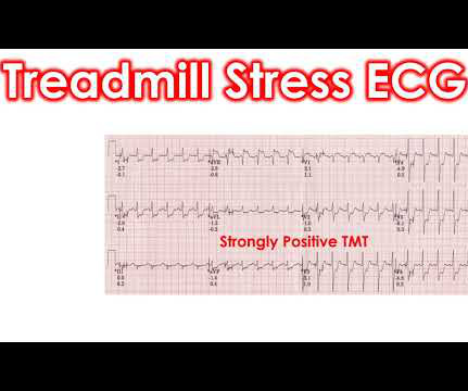
Treadmill Exercise ECG is usually done with a computerized treadmill unit which controls the motor speed of the treadmill as well as monitors the ECG. Treadmill exercise test ECG series starts with the pretest ECG and recordings in every stage of exercise and recovery phase. But the ST segment is down sloping in inferior leads.
In this patient's case, the RV ischemia manifested as dramatic anterior hyperacute T waves. This degree of STE is a bit atypical for LAD ischemia. Remember that the RV is the most anterior chamber. Here is a transverse image of a CT showing this. A few clues that might have suggested this are: There is marked STE in V1.
This usually represents posterior OMI, but in tachycardia and especially after cardiac arrest, this could simply be demand ischemia, residual subendocardial ischemia due to the low flow state of the cardiac arrest. This rules out subendocardial ischemia and is diagnostic of posterior OMI. V4-5 continue to show STD.
Additionally, it offers clinicians the flexibility to perform exercise stress testingsomething not feasible with any previously available cardiac PET tracer. The longer half-life eliminates the need for onsite tracer manufacturing so it can be ordered as a ready-to-use unit dose.
This may result in ischemia (lack of oxygen to the heart muscle), causing parts of the heart to weaken and enlarge. Exercise regularly to keep the heart strong and healthy. Coronary Artery Disease (CAD) CAD, which involves the narrowing or blockage of coronary arteries due to plaque buildup, can reduce blood flow to the heart.
We have previously shown that ablation of CaMKIIδ oxidation by CRISPR-Cas9 base editing enables the heart to recover function from otherwise severe damage following ischemia/reperfusion (IR) injury. Here, we extended this therapeutic concept toward potential clinical translation.
She underwent exercise echocardiogram in mid October where she exercised for nearly 7 minutes on the standard Bruce protocol and had typical anginal pain and shortness of breath. Time 17 minutes Not much different One month earlier This is Left Bundle Branch Block (LBBB) without any sign of ischemia. link] Shvilkin et al.
Reference : Apart from the heavily quoted classics of COURAGE, BARI-2D, ISCHEMIA, ORBITA 1 etc. Regular exercise equivalent to PCI (ESC 2009).Will Please note ORBITA -2 is not an antidote to ORBITA-1) ,Read this 1.AVERT AVERT study :Atorvastatin equals PCI.2.Regular Will try to get the link for this soon.
We are told that the Stress Echo that was performed showed objective evidence of inducible ischemia ( confirmed apparently by both wall motion abnormalities and ECG changes ). Was this objective evidence of inducible ischemia accompanied by chest pain? Was this objective evidence of inducible ischemia accompanied by chest pain?
If you have narrowing in blood vessels which only stop blood from getting through at times of stress or exercise then an ECG or an ECHO at rest may be completely normal despite there being a problem. These are therefore not looking for coronary disease but instead ischemia heart disease. Here the patient is given some dye.
Furthermore, she denies any hydration since conclusion of exercise. There is broad subendocardial ischemia as demonstrated by STE aVR with concomitant STD that almost appears appropriately maximal in Leads II and V5. There is LBBB-like morphology with persistent patterns of subendocardial ischemia.
ET Main Tent (Hall B1) This session offers more insights from key clinical trials presented at ACC.24 24 and find out what it all means for your patients.
And superimposed subendocardial ischemia pattern, of course. Before continuing flecainide, he had me get on a treadmill at full dose and at full exercise (18 minutes) and measured the QRS to be certain that the QRS did not lengthen at all. She was otherwise very stable during this rhythm. Coincidence?).
The ECG in Figure-1 was obtained from a previously healthy middle-aged man — who while performing his regular exercise routine, developed "slight" chest discomfort and "palpitations". ie, Severe subendocardial ischemia from sustained VT in a patient severe apical cardiomyopathy resulted in a peak troponin >31,000 ng/L in today's case ).
Learning Points: Ectopic atrial rhythm can produce atrial repolarization findings that can be confused for acute ischemia, STEMI, or OMI. If you can safely and easily increase the patient's heart rate, you can convert the patient to sinus and repeat the ECG to see if the atrial repolarization wave was the cause of the concern for ischemia.
After six weeks, there was no difference in angina symptoms or exercise capacity between these two groups. Subscribe now 1 ISCHEMIA Research Group. Most importantly, patients were unaware if they had received a stent during their procedure. The result of this trial sent shockwaves through the world of cardiology. N Engl J Med.
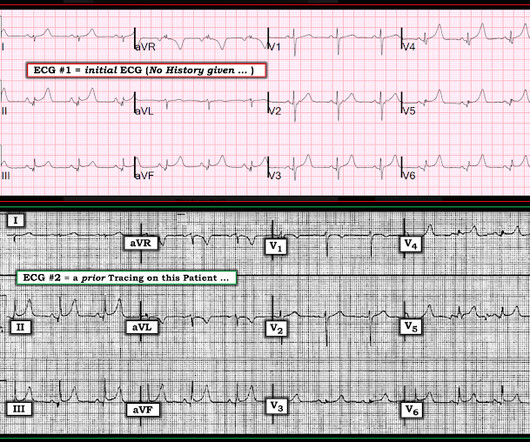
It is interesting to speculate on WHY these differences might exist between ECG #1 and ECG #2: It could represent ischemia. Since the reason for obtaining ECG #1 was “ICD malfunction” — perhaps there was a sustained arrhythmia — and T wave changes reflect a “memory effect”?
Additionally, the heart’s high demand for oxygen makes it particularly vulnerable to conditions like ischemia (reduced blood flow), which can weaken or damage the heart muscle if left untreated. Common Problems in Coronary Circulation Understanding coronary circulation helps shed light on common heart conditions.
They include myocardial ischemia, acute pericarditis, pulmonary embolism, external compression due to mass over the right ventricular outflow tract region, and metabolic disorders like hyper or hypokalemia and hypercalcemia. These are the conditions which have to be considered or excluded as they can sometimes manifest Brugada pattern on ECG.
Next day, a stress echo was done: The exercise stress echocardiogram is normal. This ST-T wave appearance in the lateral chest leads of ECG #2 is consistent with L V “ S train” vs ischemia. Normal estimated left ventricular ejection fraction improved with stress. No wall motion abnormality at rest.
Whereas at low to moderate degrees of exercise, the risk of developing AFib in younger athletic individuals is reduced — there appears to be a “threshold” for exercise intensity with longterm endurance training, beyond which the risk of developing AFib paradoxically increases!
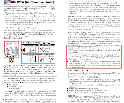
As discussed in ECG Blog #363 — this rare genetic disorder almost always presents in association with emotional stress or with exercise (ie, CPVT is usually "induced" by catecholamine discharge ). ECG Blog #157 — Can you diagnose ischemia and/or infarction when there is WPW?
When the differential is LAD occlusion versus Normal Variant, use the formula as an assistive tool – only exercise extreme caution when reclassifying what you believe to be LAD occlusion as Normal Variant. It’s important to stress the presence of a normal QRS (i.e., Benign ST/T configuration can be dramatic, and many times frightening.
This approach also reduces death from heart disease and heart attacks by 41% compared to conventional approaches such as exercise stress testing 2. 5 ISCHEMIA Research Group. The use of CTCA is often the only test that is required, therefore, avoiding further invasive testing. Eur Heart J. 2020 Jan 14;41(3):407-477. N Engl J Med.
Suspect an idiopathic form of VT when a younger adult without known coronary or structural heart disease develops a regular WCT ( W ide- C omplex T achycardia ) rhythm during exercise or other strenuous effort — and despite this, seems to tolerate the WCT rhythm surprisingly well.
This is one case where it made a difference: Right Ventricular MI seen on ECG helps Angiographer to find Culprit Lesion Nevertheless, it is sometimes a fun academic exercise to try to predict the infarct artery: An elderly patient had onset of chest pain one hour prior. He called 911. Here is the prehospital ECG. What do you think?
Sent by anonymous, written by Pendell Meyers A male in his teens presented with complaints of chest discomfort and dyspnea beginning while exercising but without obvious injury. He immediately stopped exercising and symptoms started to improve.
Such findings would normally suggest primary ischemia with concomitant surveillance of coronary occlusion, but these ST/T changes might very well be secondary to the Escape mechanism at hand. Lead V2 shows RR’ QRS configuration, and although ST depression is otherwise expected here, the discordance is a bit excessive.
We investigated the prognostic value of this incongruity, considering both known atherosclerosis and myocardial ischemia.Methods:In a retrospective analysis, we examined 111 patients (mean age: 64±12 years, 58% women) who underwent exercise stress echocardiography, with recent carotid artery and coronary evaluation.
Background and Purpose:Inflammation plays a significant role in acute and chronic cerebral ischemia. We aimed to determine if C-RIC and physical exercise (EXR) attenuated the increase in the NLRP3 inflammasome and related cytokines in the BCAS model.Methods:Microcoil-induced BCAS was used for chronic hypoperfusion, a model for VCID.
ETT ( E xercise T readmill T est ) — to see what happens to this patient's heart rate and how he handles progressively increasing levels of exercise. ETT — excellent level of activity and heart rate response to exercise. No evidence of ischemia. Negative family history for sudden death or arrhythmia. Echo — completely normal.
In MSIMI (Mental Stress-induced Myocardial Ischemia) studies , mental stress activities like public speaking were evaluated for their impact on ischemia, measured via myocardial SPECT and vascular function (microvascular function, endothelial function). Moreover, women under 50 years old are four times more likely to experience MSIMI.
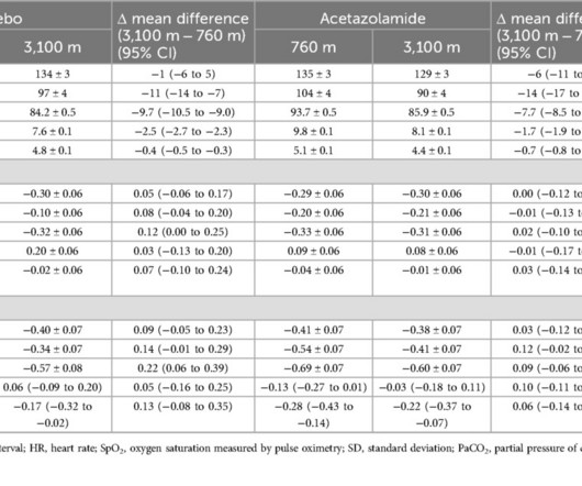
Exercise electrocardiograms were recorded at the National Center of Internal Medicine and Cardiology, Bishkek (760m) and on the day of arrival at the Tuja Ashu high-altitude clinic (3,100m), Kyrgyzstan. The mean difference (95% CI) in STE between post-peak exercise between 3,100m and 760m was 0.22mm (0.06 to 0.39) and 0.09mm (0.06
We organize all of the trending information in your field so you don't have to. Join thousands of users and stay up to date on the latest articles your peers are reading.
You know about us, now we want to get to know you!
Let's personalize your content
Let's get even more personalized
We recognize your account from another site in our network, please click 'Send Email' below to continue with verifying your account and setting a password.


Let's personalize your content