This site uses cookies to improve your experience. To help us insure we adhere to various privacy regulations, please select your country/region of residence. If you do not select a country, we will assume you are from the United States. Select your Cookie Settings or view our Privacy Policy and Terms of Use.
Cookie Settings
Cookies and similar technologies are used on this website for proper function of the website, for tracking performance analytics and for marketing purposes. We and some of our third-party providers may use cookie data for various purposes. Please review the cookie settings below and choose your preference.
Used for the proper function of the website
Used for monitoring website traffic and interactions
Cookie Settings
Cookies and similar technologies are used on this website for proper function of the website, for tracking performance analytics and for marketing purposes. We and some of our third-party providers may use cookie data for various purposes. Please review the cookie settings below and choose your preference.
Strictly Necessary: Used for the proper function of the website
Performance/Analytics: Used for monitoring website traffic and interactions
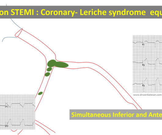
True bifurcation STEMI with static thrombus (Carinal trapping of thrombus ,Coronary Lerish sydrome ) 4. EmbolicSTEMI with showers of emboli into both LCX and LAD Simultaneous or sequential Anterior and Inferior STEMI 5. Wrap around LAD true Global MI 2. RCA-dependent LAD circulation through collaterals 3.
for those of you who do not do Emergency Medicine, ECGs are handed to us without any clinical context) The ECG was read simply as "No STEMI." He was started on a heparin drip and CTA of the chest was ordered to rule out pulmonary embolism. Unfortunately, there was a long wait and the patient left before being seen by a provider.
This ECG is diagnostic of anterior STEMI. The distal inferior apical LAD was cut off by distal embolization from LAD culprit. The QRS is at least as important as the ST segment in diagnosing STEMI It has been constant since then. He looked ill and diaphoretic. BP was 160. And the Cath lab was activated immediately.
In this ECG Cases blog we look at 10 cases of patients with chest pain, including false positive STEMI, false negative STEMI, and other causes to help hone your ECG interpretation skills in time-sensitive cases where those very ECG skills might save a life.
We discover that for STEMI/OMI vs subendocardial ischemia, we should look for STEMI(-)OMI, subacute OMI, and OMI in the presence of LBBB and RBBB, and consider the differential for diffuse ST depression with reciprocal ST elevation in aVR.
Bi-phasic scan showed no dissection or pulmonary embolism. Take home messages: 1- In STEMI/NSTEMI paradigm you search for STE on ECG. If this patient was managed according to the STEMI/NSTEMI paradigm (although he would be a candidate for early invasive treatment), he would probably be taken to the cath lab hours later.
The following ECG was recorded: There is an obvious acute inferior STEMI. Whenever there is inferior STEMI, one should think about Right Ventricular STEMI (RVMI). As 85% of inferior STEMI are due to RCA occlusion [the rest due to occlusion of a "dominant" circumflex (i.e., and STE in lead III > STE in lead II.
The prehospital and ED computer interpretation was inferior STEMI: There’s normal sinus rhythm, first degree AV block and RBBB, normal axis and normal voltages. Smith comment: before reading anything else, this case screamed pulmonary embolism to me. The prehospital, ED computer, and final cardiology interpretation was STEMI negative.
We discuss current and emerging techniques to extract or disperse thrombi, aiming to reduce downstream embolization, microvascular obstruction and myocardial injury. However, innovations in stroke intervention have sparked renewed interest in thrombus modification approaches.
This certainly looks like an anterior STEMI (proximal LAD occlusion), with STE and hyperacute T-waves (HATW) in V2-V6 and I and aVL. How do you explain the anterior STEMI(+)OMI immediately after ROSC evolving into posterior OMI 30 minutes later? This caused a type 2 anterior STEMI.
It makes pulmonary embolism (PE) very likely. Although most cardiac arrest from MI is due to ventricular fibrillation, some is due to high grade AV block, and so this could indeed be due to large acute STEMI. LV anterior STEMI does not give maximal ST elevation in V1. So this ECG is typical of right ventricular (RV) STEMI.
The conventional machine algorithm interpreted this ECG as STEMI. In patients with narrow QRS ( not this patient), this pattern is highly suggestive of acute pulmonary embolism. See this post of RV MI with both McConnell sign and "D" sign: Inferior and Posterior STEMI. When EMS found her, she was dyspneic and diaphoretic.
This ECG is highly concerning for LAD occlusion despite it not showing a STEMI criteria. You can find the variables used to calculate the value on MD calc here: [link] Utilizing Dr. Smith’s Subtle Anterior STEMI Calculator (4-Variable), the value is greater than 18.2 which is concerning for LAD occlusion.
Notice on the right side of the image how the algorithm correctly measures STE sufficient in V1 and V2 to meet STEMI criteria in a man older than age 40. As most would agree, this ECG shows highly specific findings of anterolateral OMI, even with STEMI criteria in this case. Thus, this is obvious STEMI(+) OMI until proven otherwise.
Transient and partial thrombosis at the site of a non-obstructive plaque with subsequent spontaneous fibrinolysis and distal embolization may be one of the mechanisms responsible for the occurrence of MINOCA. This has resulted in an under-representation of STEMI MINOCA patients in the literature. From Gue at al. Circulation.
Smith interpretation: This is highly likely to be due to extreme right heart strain and is nearly diagnostic of pulmonary embolism. She was diagnosed with a Non-STEMI and kept overnight for a next day angiogram. Medics recorded the above ECG and called a STEMI alert. It is of course pulmonary embolism.
Is this an anterior STEMI with LBBB? Explanation : The patient had a worrisome history: 59 yo with significant substernal chest pressure, so his pretest probability of MI (and even of STEMI) is reasonably high. Additionally, appropriate discordance is common in NonSTEMI, but very unusual in coronary occlusion (STEMI).
This meets "STEMI criteria" However, there is very high voltage, with a very deep S-wave in V2 and tall R-wave in V4. The morphology is not right for STEMI. My interpretation: LVH with secondary ST-T abnormalities, exaggerated by stress, not a STEMI. This is very good evidence that the ST elevation is not due to STEMI.
The "criteria" for posterior STEMI are 0.5 Is it STEMI or NonSTEMI? The patient had been on a long drive, suggesting possible pulmonary embolism (this was unlikely given absence of tachyardia, hypoxia, or any other feature of PE), so we sent a d dimer. The troponin I returned at 4.1 mm STE in one lead. This includes: 1.
In patients with nonvalvular A fib, the majority of embolic strokes are caused by thrombi development in the left atrial appendage. Patients with documented STEMI, left ventricular thrombus, mechanical mitral or aortic valve replacement were excluded. A fib is a well-established risk factor for ischemic strokes.
There is inferior STEMI. He took another look and realized that the culprit was indeed in the proximal RCA and that the thrombus had embolized distally. For those who don't have time to watch a video, you'll have to read the ECG as shown on this still frame because I lost it and cannot post it. There was no right sided ECG.
It is uncommon in the age of reperfusion therapy, as most STEMI get treated reasonably early, before transmural infarct. LV aneurysm puts them at risk for a mural thrombus, which puts them at risk for embolism, especially embolic stroke. Most STEMI peak at over 10 ng/mL; most NonSTEMI at less than 10 ng/mL.
The morphology of V2-V4 is very specific in my experience for acute right heart strain (which has many potential etiologies, but none more common and important in EM than acute pulmonary embolism). CT angiogram showed extensive saddle pulmonary embolism. He had multiple cardiac arrests with ROSC regained each time. This is a quiz.
Without seeing the patient, my interpretation of the first ECG was: likely normal variant ST-elevation (early repolarization), with a small possibility of pericarditis, and almost no possibility of acute coronary occlusion (STEMI). and therefore highly unlikely to be STEMI. Does subsegmental pulmonary embolism matter?
In this ECG Cases blog, Jesse McLaren and Rajiv Thavanathan explore how ECG and POCUS complement each other for patients presenting to the emergency department with shortness of breath or chest pain. They explain complementary diagnostic insights into pericardial effusion and cardiac tamponade, occlusion MI and RV strain.
This is a troponin I level that is almost exclusively seen in STEMI. So this is either a case of MINOCA, or a case of Type II STEMI. If the arrest had another etiology (such as old scar), and the ST elevation is due to severe shock, then it is a type II STEMI. I believe the latter (type II STEMI) is most likely.
Later, she developed chest pain again, and had this ECG recorded: Obvious Anterior OMI that is also a STEMI Coronary angiogram- --Right dominant coronary artery system --The left main artery was normal in appearance and free of obstructive disease. --The Thus, Wellens' syndrome should be thought of as a transient OMI or transient STEMI.
This is of course diagnostic of an acute coronary occlusion MI (OMI) that also meets STEMI criteria. However, by the time of the angiogram it had embolized distally, and had only done so after the right sided ECG was recorded. This prehospital ECG was recorded: Here are limb leads: Here are precordial leads: Diagnosis?
Transient and partial thrombosis at the site of a non-obstructive plaque with subsequent spontaneous fibrinolysis and distal embolization may be one of the mechanisms responsible for the occurrence of MINOCA. This has resulted in an under-representation of STEMI MINOCA patients in the literature. From Gue at al. Circulation.
50% of LAD STEMIs do not have reciprocal findings in inferior leads, and many LAD OMIs instead have STE and/or HATWs in inferior leads instead. The ECG easily meets STEMI criteria in all leads V2-V6, as well. CT angiogram chest: no aortic dissection or pulmonary embolism. 24 yo woman with chest pain: Is this STEMI?
The commonest causes of MINOCA include: atherosclerotic causes such as plaque rupture or erosion with spontaneous thrombolysis, and non-atherosclerotic causes such as coronary vasospasm (sometimes called variant angina or Prinzmetal's angina), coronary embolism or thrombosis, possibly microvascular dysfunction.
In a series of 18 patients with COVID and ST elevation, 8 were diagnosed with STEMI, 6 of whom had an angiogram and it showed obstructive coronary disease. 12 All STEMI patients had very high cTn typical of STEMI (cTnT > 1.0 Smith ) — the overall impression was that ECG #1 did not suggest findings suggestive of OMI.
Both of these are very suggestive of " No-Reflow ," or poor microvascular reperfusion due to downstream embolization of microscopic platelet-fibrin aggregates. cm diameter in the apex The presence of thrombus led the clinicians to state that this was a "late presentation STEMI." LV Thrombus , 1.5 0 0 1 95 544 MMRF 4 1 638 14.0
ECG read as: "Shows T wave inversions in the inferior leads and less than 1mm STE in V2, without STEMI criteria." CT pulmonary angiogram was negative for pulmonary embolism. All very very subtle. So the patient was placed back in the waiting room like many others. Aspirin was given. Second troponin T resulted at 1,318 ng/L.
You may see a filling defect in distal LAD, most probably due to an embolization from proximal lesion. The "occlusion pattern" on ECG may change during the course of evolution ( in today's case — from a proximal LAD OMI to a more distal LAD OMI pattern, presumably due to embolization from a proximal lesion ).
In this ECG Cases blog we look at 6 patients who presented with cardiorespiratory symptoms, possibly from COVID and illustrate the dangers of anchoring, being hypervigilant for cardiovascular complications, and why testing for COVID in patients being admitted for ACS is important.
In this ECG Cases blog we review 10 cases of possible artifact, lead reversal and lead misplacement. Can you spot the abnormalities and avoid the misdiagnosis? The post ECG Cases 29 Misdiagnosis from Lead Misplacement, Artifact and Lead Reversal appeared first on Emergency Medicine Cases.
Patients with pulmonary embolism or aortic dissection who have normal variant ST elevation are at high risk of being diagnosed with pericarditis when what they have is far more serious!! normal variant, not pericarditis) A Young Man with Sharp Chest pain (normal variant, not pericarditis) 24 yo woman with chest pain: Is this STEMI?
The limb leads have been removed because there was no ST elevation in those leads, the QRS complexes have been obscured because this is irrelevant to STEMI criteria, and red lines have been added to measure ST segment elevation. But STEMI criteria ignore all this and look at ST segments in isolation.
The emergency medicine physician documented, "His initial EKG is riddled with artifact and difficult to interpret but does not look like a STEMI." The ECG remains positive for STEMI by GE. Even if it is not atherosclerotic, young people can have embolic OMIs. In fact, even the GE algorithm got this one (partially) right.
Note: the 2022 ACC Expert consensus Chest pain guidelines state that "posterior STEMI-Equivalent" is a sign of acute coronary occlusion. 2/3 of STEMI have a peak 4th generation troponin I greater than 10.0 Comment: The first ECG is diagnostic of OMI that does not meet STEMI criteria. NSTEMI-OMI").
Third, a slow motion segment showing delayed, brisk filling of the PDA due to dislodgment of a thrombus from contrast injection and distal embolization. A distal RCA lesion ( blue arrow ), Delayed brisk filling of an initially occluded PDA due to a thrombus dislodged during injection which embolized distally.
His initial high sensitivity troponin I returned at 1300 ng/L and given that his cardiac workup was otherwise unremarkable, a CT was obtained to evaluate for pulmonary embolism and aortic aneurysm or dissection but this too was unrevealing. Another EKG was also obtained. ECG at time 82 minutes: What do you think?
The cath lab was deactivated by cardiologist on arrival at ED because it was "not a STEMI". No pulmonary embolism is identified. Pt received 324 ASA and 2 sprays of nitro with improvement. Cath lab was activated by EMS and transported emergent." Further events: The patient continued to have pain and an NTG drip was started.
We organize all of the trending information in your field so you don't have to. Join thousands of users and stay up to date on the latest articles your peers are reading.
You know about us, now we want to get to know you!
Let's personalize your content
Let's get even more personalized
We recognize your account from another site in our network, please click 'Send Email' below to continue with verifying your account and setting a password.


Let's personalize your content