This site uses cookies to improve your experience. To help us insure we adhere to various privacy regulations, please select your country/region of residence. If you do not select a country, we will assume you are from the United States. Select your Cookie Settings or view our Privacy Policy and Terms of Use.
Cookie Settings
Cookies and similar technologies are used on this website for proper function of the website, for tracking performance analytics and for marketing purposes. We and some of our third-party providers may use cookie data for various purposes. Please review the cookie settings below and choose your preference.
Used for the proper function of the website
Used for monitoring website traffic and interactions
Cookie Settings
Cookies and similar technologies are used on this website for proper function of the website, for tracking performance analytics and for marketing purposes. We and some of our third-party providers may use cookie data for various purposes. Please review the cookie settings below and choose your preference.
Strictly Necessary: Used for the proper function of the website
Performance/Analytics: Used for monitoring website traffic and interactions
In this phenomenon, a thrombus forms within the lumen of the stent graft component of the frozen elephant trunk prosthesis and puts the patient at risk for downstream embolization with visceral or lower limb ischemia. Therefore, the presence of ILT is associated with increased short-term mortality and morbidity.
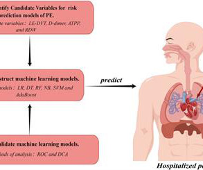
Baseline data were analyzed using univariate analysis, and potential independent riskfactors associated with PE were further identified through univariate and multivariate logistic regression analysis. Participants were randomly divided into a training group (70%) and a validation group (30%).
Genetic protein S (PS) deficiency caused by PROS1 gene mutation is an important riskfactor for hereditary thrombophilia.Case introductionIn this case, we report a 28-year-old male patient who developed a severe pulmonary embolism during his visit. Ultrasound showed no thrombosis in the veins of both lower limbs.
Inferior vena cava (IVC) agenesis is a rare congenital anomaly that has been implicated in up to 5% of unprovoked deep vein thrombosis (DVT) cases in young men under 30 years old. A hypercoagulability workup was positive for a heterozygous Factor V Leiden (FVL) mutation, an additional thrombophilic riskfactor.
MINOCA may be due to: coronary spasm, coronary microvascular dysfunction, plaque disruption, spontaneous coronary thrombosis/emboli , and coronary dissection; myocardial disorders, including myocarditis, takotsubo cardiomyopathy, and other cardiomyopathies. This is in spite of the known proclivity of tighter stenoses to thrombose.
However, it subjects patients to lifelong warfarin therapy after MHVR, with the attendant risk of bleeding and thrombosis. The percentage of time in the therapeutic range (TTR) was used as the primary outcome while bleeding, thrombosis, and other events were the secondary outcomes.Results:A total of 721 patients were enrolled.
The commonest causes of MINOCA include: atherosclerotic causes such as plaque rupture or erosion with spontaneous thrombolysis, and non-atherosclerotic causes such as coronary vasospasm (sometimes called variant angina or Prinzmetal's angina), coronary embolism or thrombosis, possibly microvascular dysfunction.
pulmonary embolism, sepsis, etc.), Coronary thrombosis or embolism can result in MINOCA, either with or without a hypercoagulable state. If there is any evidence of atherosclerosis, modifiable CAD riskfactors should be treated aggressively. 2) overlooked obstructive coronary disease (e.g., myocarditis).
CT angiogram chest: no aortic dissection or pulmonary embolism. Smith Major Learning Point: The worst riskfactor for a bad outcome in OMI is young age because cardiologists cannot believe that a young person can have an OMI. No further troponins were measured. Serial chest xrays: progressive bilateral pulmonary edema.
Introduction:COVID-19 infection has thus emerged to be a new riskfactor for Cerebral Venous Thrombosis (CVT). Acute ischemic stroke (AIS), intracerebral hemorrhage (ICH), subarachnoid hemorrhage (SAH), epilepsy, deep vein thrombosis and pulmonary embolism were secondary outcomes. vs 5.5%; HR=2.16; 95% CI=1.23-3.64
We organize all of the trending information in your field so you don't have to. Join thousands of users and stay up to date on the latest articles your peers are reading.
You know about us, now we want to get to know you!
Let's personalize your content
Let's get even more personalized
We recognize your account from another site in our network, please click 'Send Email' below to continue with verifying your account and setting a password.


Let's personalize your content